 Explore
 Validate
 Learn
Learn Flow cytometry
Antibody data
- Antibody Data
- Antigen structure
- References [5]
- Comments [0]
- Validations
- Flow cytometry [2]
- Other assay [3]
Submit
Validation data
Reference
Comment
Report error
- Product number
- 12-5841-82 - Provider product page

- Provider
- Invitrogen Antibodies
- Product name
- OCT3/4 Monoclonal Antibody (EM92), PE, eBioscience™
- Antibody type
- Monoclonal
- Antigen
- Other
- Description
- Description: The EM92 monoclonal antibody reacts with mouse and human Oct3/4, encoded by the Pou5F1 gene. Oct3/4 is a POU domain-containing transcription factor that is critical for maintaining embryonic stem (ES) and induced pluripotent stem (iPS) cells in a pluripotent state, and is expressed by ES, embryonic germ cells and embryonic carcinoma cell lines. In cells of the inner cell mass (ICM), reduction of Oct3/4 expression causes dedifferentiation to trophoectoderm, whereas increased expression results in differentiation to mesoderm and primitive endoderm. Oct3/4 regulates the expression of several genes, including FGF-4, UTF1, Sox2, Fbx15, Rex1 and osteopontin through distinct mechanisms. Furthermore, Oct3/4 frequently acts synergistically with Sox2 to regulate target gene expression, as is the case with FGF-4. It has been demonstrated that Oct3/4 expression in ES cells can be negatively regulated by either treatment with retinoic acid, or by removal of leukemia-inhibitory factor (LIF). Applications Reported: This EM92 antibody has been reported for use in intracellular staining followed by flow cytometric analysis. Applications Tested: This EM92 antibody has been tested by intracellular staining and flow cytometric analysis of F9 embryonal carcinoma cells using the Foxp3/Transcription Factor Staining Buffer Set (Product # 00-5523-00) and protocol. Please see BestProtocols® for Staining Protocol (refer to Protocol B: One-step protocol for intracellular (nuclear) proteins). This antibody can be used at less than or equal to 0.5 µg per test. A test is defined as the amount (µg) of antibody that will stain a cell sample in a final volume of 100 µL. Cell number should be determined empirically but can range from 10^5 to 10^8 cells/test. It is recommended that the antibody be carefully titrated for optimal performance in the assay of interest. Excitation: 488-561 nm; Emission: 578 nm; Laser: Blue Laser, Green Laser, Yellow-Green Laser. Filtration: 0.2 µm post-manufacturing filtered.
- Reactivity
- Human, Mouse
- Host
- Rat
- Conjugate
- Yellow dye
- Isotype
- IgG
- Antibody clone number
- EM92
- Vial size
- 100 µg
- Concentration
- 0.2 mg/mL
- Storage
- 4°C, store in dark, DO NOT FREEZE!
Submitted references CBP-mediated Wnt3a/β-catenin signaling promotes cervical oncogenesis initiated by Piwil2.
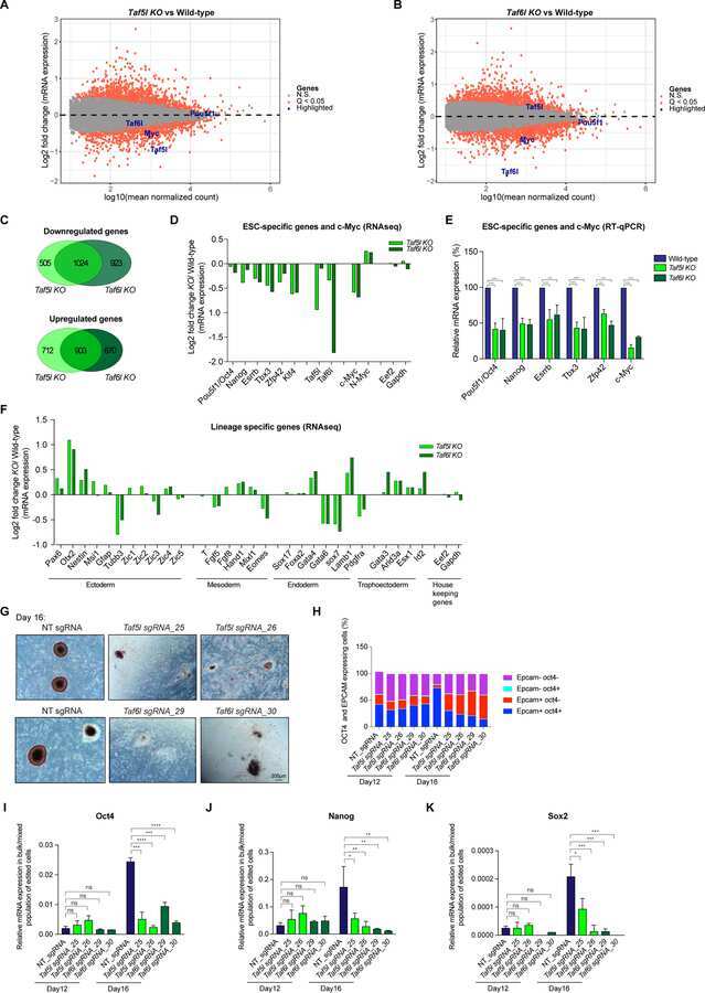
TAF5L and TAF6L Maintain Self-Renewal of Embryonic Stem Cells via the MYC Regulatory Network.
The HUSH complex cooperates with TRIM28 to repress young retrotransposons and new genes.
Generation of iPS Cells from Human Peripheral Blood Mononuclear Cells Using Episomal Vectors.
Highly upregulated Lhx2 in the Foxn1-/- nude mouse phenotype reflects a dysregulated and expanded epidermal stem cell niche.
Feng D, Yan K, Liang H, Liang J, Wang W, Yu H, Zhou Y, Zhao W, Dong Z, Ling B
Neoplasia (New York, N.Y.) 2021 Jan;23(1):1-11
Neoplasia (New York, N.Y.) 2021 Jan;23(1):1-11
TAF5L and TAF6L Maintain Self-Renewal of Embryonic Stem Cells via the MYC Regulatory Network.
Seruggia D, Oti M, Tripathi P, Canver MC, LeBlanc L, Di Giammartino DC, Bullen MJ, Nefzger CM, Sun YBY, Farouni R, Polo JM, Pinello L, Apostolou E, Kim J, Orkin SH, Das PP
Molecular cell 2019 Jun 20;74(6):1148-1163.e7
Molecular cell 2019 Jun 20;74(6):1148-1163.e7
The HUSH complex cooperates with TRIM28 to repress young retrotransposons and new genes.
Robbez-Masson L, Tie CHC, Conde L, Tunbak H, Husovsky C, Tchasovnikarova IA, Timms RT, Herrero J, Lehner PJ, Rowe HM
Genome research 2018 Jun;28(6):836-845
Genome research 2018 Jun;28(6):836-845
Generation of iPS Cells from Human Peripheral Blood Mononuclear Cells Using Episomal Vectors.
Su RJ, Neises A, Zhang XB
Methods in molecular biology (Clifton, N.J.) 2016;1357:57-69
Methods in molecular biology (Clifton, N.J.) 2016;1357:57-69
Highly upregulated Lhx2 in the Foxn1-/- nude mouse phenotype reflects a dysregulated and expanded epidermal stem cell niche.
Bohr S, Patel SJ, Vasko R, Shen K, Huang G, Yarmush ML, Berthiaume F
PloS one 2013;8(5):e64223
PloS one 2013;8(5):e64223
No comments: Submit comment
Supportive validation
- Submitted by
- Invitrogen Antibodies (provider)
- Main image

- Experimental details
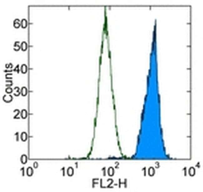
- Staining of F9 embryonal carcinoma cell line with 0.25 µg of Rat IgG2a K Isotype Control PE (Product # 12-4321-80) (open histogram) or 0.25 µg of Anti-Mouse OCT3/4 PE (filled histogram). Staining was carried out using the Foxp3 Staining Buffer Set (00-5523). Total cells were used for analysis.
- Conjugate
- Yellow dye
- Submitted by
- Invitrogen Antibodies (provider)
- Main image

- Experimental details
- Staining of F9 embryonal carcinoma cell line with 0.25 µg of Rat IgG2a K Isotype Control PE (Product # 12-4321-80) (open histogram) or 0.25 µg of Anti-Mouse OCT3/4 PE (filled histogram). Staining was carried out using the Foxp3 Staining Buffer Set (00-5523). Total cells were used for analysis.

Supportive validation
- Submitted by
- Invitrogen Antibodies (provider)
- Main image
- Experimental details
- NULL
- Conjugate
- Yellow dye
- Submitted by
- Invitrogen Antibodies (provider)
- Main image
- Experimental details
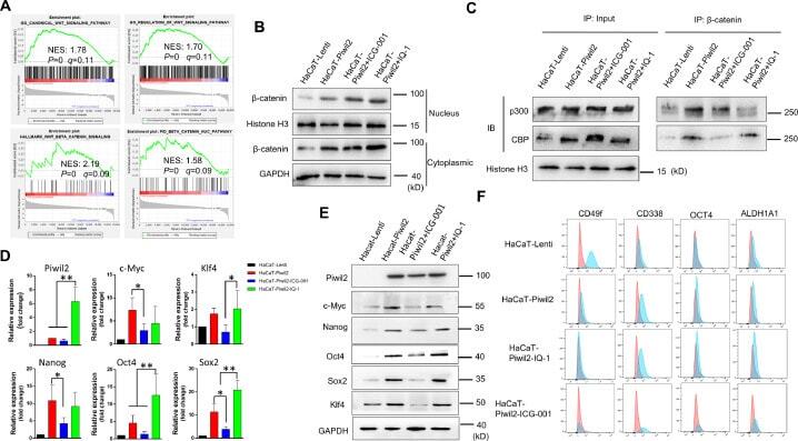
- Figure 4 CBP/beta-catenin promotes the maintenance of stem cell reprogramming by Piwil2. (A) GSEA plot showing significant enrichment of the Wnt/beta-catenin signaling activation modules in HaCaT-Piwil2 cells. (B) Immunoblotting of beta-catenin translocated into the nucleus in HaCaT-Piwil2 cells after treatment with 20 uM ICG-001 or IQ-1 for 24 h. (C) Nuclear lysates from HaCaT-Piwil2 cells treated with 20 uM ICG-001, 20 uM IQ-1, or DMSO were coimmunoprecipitated with antisera to beta-catenin and immunoblotted for CBP and p300. (D and E) The expression of Piwil2 and the ""reprogramming"" factors c-Myc, Nanog, Oct4, Sox2 , and Klf4 was determined by real-time PCR and immunoblotting in HaCaT-Piwil2 cells treated with 20 uM ICG-001, 20 uM IQ-1, or DMSO for 24 h. (F) The proportion of CD49f-, CD338-, OCT4-, and ALDHA1-positive cells, determined by flow cytometry in HaCaT-Piwil2 cells treated with 20 uM ICG-001, 20 uM IQ-1, or DMSO for 24 h. The data are presented as the mean +- SD. * P < 0.05 and ** P < 0.01 by Student's t test. Figure 4
- Conjugate
- Yellow dye
- Submitted by
- Invitrogen Antibodies (provider)
- Main image
- Experimental details
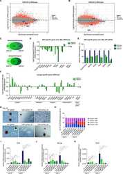
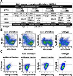
- Figure 4 As Oct3/4+ population is expanded; CD49f+, CD34+ and CD200+ populations are reduced in epithelial isolates from newborn skin of the Foxn1 -/- phenotype vs. wild type. (A) Flow cytometry summary table listing fractions positive for markers of progeny within epithelial isolates from newborns (%+-SD, n>6). Distinct differences between the Foxn1 -/- phenotype and wild type are demonstrated by comparative sample flow cytometry data gated for (B) CD49f, CD34, CD200 and (C) Oct3/4 positive epithelial fractions.
- Conjugate
- Yellow dye
