Explore
 Validate
 Learn
Learn Western blot
 Immunoprecipitation
Antibody data
- Antibody Data
- Antigen structure
- References [6]
- Comments [0]
- Validations
- Western blot [5]
- Immunocytochemistry [2]
- Immunohistochemistry [2]
Submit
Validation data
Reference
Comment
Report error
- Product number
- GTX101566 - Provider product page

- Provider
- GeneTex
- Proper citation
- GeneTex Cat#GTX101566, RRID:AB_11177712
- Product name
- Galectin 1 antibody
- Antibody type
- Polyclonal
- Reactivity
- Human, Mouse, Rat
- Host
- Rabbit
Submitted references Upregulation of LGALS1 is associated with oral cancer metastasis.
Expression of Galectins-1 and Galectin-3 in Stomach and Colorectal Cancer with Tissue Eosinophilia.
A potential role of galectin-1 in promoting mouse trophoblast stem cell differentiation.
Comparative Proteomic and Transcriptomic Analysis of Follistatin-Induced Skeletal Muscle Hypertrophy.
Resources for the Comprehensive Discovery of Functional RNA Elements.
Secretome protein signature of human pancreatic cancer stem-like cells.
Li JM, Tseng CW, Lin CC, Law CH, Chien YA, Kuo WH, Chou HC, Wang WC, Chan HL
Therapeutic advances in medical oncology 2018;10:1758835918794622
Therapeutic advances in medical oncology 2018;10:1758835918794622
Expression of Galectins-1 and Galectin-3 in Stomach and Colorectal Cancer with Tissue Eosinophilia.
Kolobovnikova YV, Dmitrieva AI, Yankovich KI, Vasil'eva OA, Purlik IL, Poletika VS, Novitskii VV, Urazova OI
Bulletin of experimental biology and medicine 2018 Jun;165(2):256-258
Bulletin of experimental biology and medicine 2018 Jun;165(2):256-258
A potential role of galectin-1 in promoting mouse trophoblast stem cell differentiation.
You JL, Wang W, Tang MY, Ye YH, Liu AX, Zhu YM
Molecular and cellular endocrinology 2018 Jul 15;470:228-239
Molecular and cellular endocrinology 2018 Jul 15;470:228-239
Comparative Proteomic and Transcriptomic Analysis of Follistatin-Induced Skeletal Muscle Hypertrophy.
Barbé C, Bray F, Gueugneau M, Devassine S, Lause P, Tokarski C, Rolando C, Thissen JP
Journal of proteome research 2017 Oct 6;16(10):3477-3490
Journal of proteome research 2017 Oct 6;16(10):3477-3490
Resources for the Comprehensive Discovery of Functional RNA Elements.
Sundararaman B, Zhan L, Blue SM, Stanton R, Elkins K, Olson S, Wei X, Van Nostrand EL, Pratt GA, Huelga SC, Smalec BM, Wang X, Hong EL, Davidson JM, Lécuyer E, Graveley BR, Yeo GW
Molecular cell 2016 Mar 17;61(6):903-13
Molecular cell 2016 Mar 17;61(6):903-13
Secretome protein signature of human pancreatic cancer stem-like cells.
Brandi J, Dalla Pozza E, Dando I, Biondani G, Robotti E, Jenkins R, Elliott V, Park K, Marengo E, Costello E, Scarpa A, Palmieri M, Cecconi D
Journal of proteomics 2016 Mar 16;136:1-12
Journal of proteomics 2016 Mar 16;136:1-12
No comments: Submit comment
Supportive validation
- Submitted by
- GeneTex (provider)
- Main image

- Experimental details
- Sample (30 ?g of whole cell lysate) A: NCI-H929 15% SDS PAGE GTX101566 diluted at 1:10000 The HRP-conjugated anti-rabbit IgG antibody (GTX213110-01) was used to detect the primary antibody.
- Submitted by
- GeneTex (provider)
- Main image
- Experimental details
- Sample (30 ?g of whole cell lysate) A: BCL-1 15% SDS PAGE GTX101566 diluted at 1:10000 The HRP-conjugated anti-rabbit IgG antibody (GTX213110-01) was used to detect the primary antibody.
- Submitted by
- GeneTex (provider)
- Main image
- Experimental details
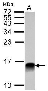
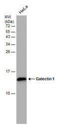
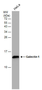
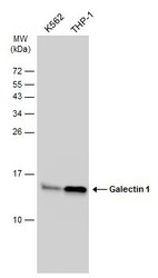
- Various whole cell extracts (30 ?g) were separated by 15% SDS-PAGE, and the membrane was blotted with Galectin 1 antibody (GTX101566) diluted at 1:10000. The HRP-conjugated anti-rabbit IgG antibody (GTX213110-01) was used to detect the primary antibody.
- Submitted by
- GeneTex (provider)
- Main image
- Experimental details
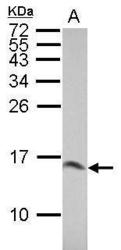
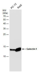
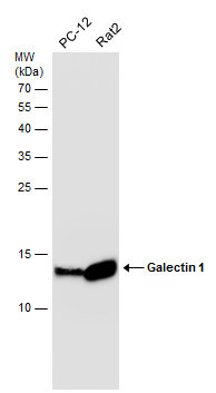
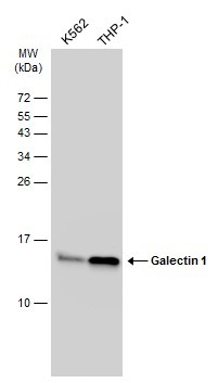
- Whole cell extract (30 ?g) was separated by 15% SDS-PAGE, and the membrane was blotted with Galectin 1 antibody (GTX101566) diluted at 1:10000. The HRP-conjugated anti-rabbit IgG antibody (GTX213110-01) was used to detect the primary antibody.
- Submitted by
- GeneTex (provider)
- Main image
- Experimental details
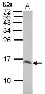
- Various whole cell extracts (30 ?g) were separated by 15% SDS-PAGE, and the membrane was blotted with Galectin 1 antibody (GTX101566) diluted at 1:2000. The HRP-conjugated anti-rabbit IgG antibody (GTX213110-01) was used to detect the primary antibody.
Supportive validation
- Submitted by
- GeneTex (provider)
- Main image
- Experimental details
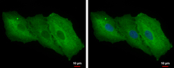
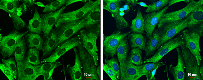
- Galectin 1 antibody detects Galectin 1 protein at cytoplasm by immunofluorescent analysis.Sample: A549 cells were fixed in 4% paraformaldehyde at RT for 15 min.Green: Galectin 1 protein stained by Galectin 1 antibody (GTX101566) diluted at 1:500.Blue: Hoechst 33342 staining.
- Submitted by
- GeneTex (provider)
- Main image
- Experimental details
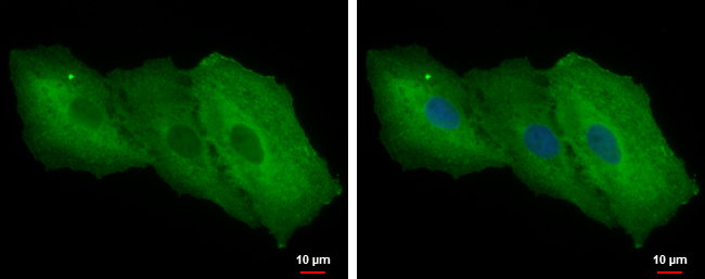
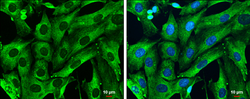
- Galectin 1 antibody detects Galectin 1 protein at cytoplasm by immunofluorescent analysis.Sample: SK-N-SH cells were fixed in 4% paraformaldehyde at RT for 15 min.Green: Galectin 1 protein stained by Galectin 1 antibody (GTX101566) diluted at 1:1000.Blue: Hoechst 33342 staining.Scale bar = 10 £gm.
Supportive validation
- Submitted by
- GeneTex (provider)
- Main image

- Experimental details
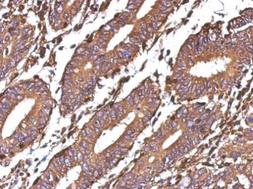
- Immunohistochemical analysis of paraffin-embedded human colon carcinoma, using Galectin 1(GTX101566) antibody at 1:500 dilution.
- Submitted by
- GeneTex (provider)
- Main image
- Experimental details
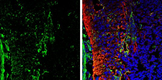
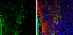
- Galectin 1 antibody detects Galectin 1 protein expression by immunohistochemical analysis.Sample: Frozen sectioned E13.5 Rat brain. Green: Galectin 1 protein stained by Galectin 1 antibody (GTX101566) diluted at 1:250.Red: beta Tubulin 3/ TUJ1, a mature neuron marker, stained by beta Tubulin 3/ TUJ1 antibody [GT11710] (GTX631836) diluted at 1:500.Blue: Fluoroshield with DAPI (GTX30920).