Explore
 Validate
 Learn
Learn Western blot
Antibody data
- Antibody Data
- Antigen structure
- References [10]
- Comments [0]
- Validations
- Western blot [2]
- Immunohistochemistry [2]
- Flow cytometry [1]
Submit
Validation data
Reference
Comment
Report error
- Product number
- AF1152 - Provider product page

- Provider
- Novus Biologicals
- Product name
- Goat Polyclonal Galectin-1 Antibody
- Antibody type
- Polyclonal
- Description
- Antigen Affinity-purified. Detects human Galectin-1 in direct ELISAs and Western blots. In direct ELISAs, approximately 40% cross-reactivity with recombinant mouse (rm) Galectin-1 is observed and less than 5% cross-reactivity with recombinant human (rh) Galectin-2, rhGalectin-3, rhGalectin-4, rhGalectin-7, rhGalectin-8, and rmGalectin-9 is observed.
- Reactivity
- Human
- Host
- Goat
- Conjugate
- Unconjugated
- Isotype
- IgG
- Vial size
- 100 ug
- Concentration
- LYOPH
- Storage
- Use a manual defrost freezer and avoid repeated freeze-thaw cycles. 12 months from date of receipt, -20 to -70 degreesC as supplied. 1 month, 2 to 8 degreesC under sterile conditions after reconstitution. 6 months, -20 to -70 degreesC under sterile conditions after reconstitution.
Submitted references Glucocorticoid receptor inhibits Müller glial galectin-1 expression via DUSP1-dependent and -independent deactivation of AP-1 signalling.
Treatment of B-cell precursor acute lymphoblastic leukemia with the Galectin-1 inhibitor PTX008.
Serum galectin-1 in patients with multiple myeloma: associations with survival, angiogenesis, and biomarkers of macrophage activation.
Galectins-1, -3, and -7 Are Prognostic Markers for Survival of Ovarian Cancer Patients.
Prototype and Chimera-Type Galectins in Placentas with Spontaneous and Recurrent Miscarriages.
Graves' disease is associated with a defective expression of the immune regulatory molecule galectin-9 in antigen-presenting dendritic cells.
Melanoma Cell Galectin-1 Ligands Functionally Correlate with Malignant Potential.
Tonsil-derived mesenchymal stem cells alleviate concanavalin A-induced acute liver injury.
Galectin-1-mediated cell death is increased by CD30-induced signaling in anaplastic large cell lymphoma cells but not in Hodgkin lymphoma cells.
Sialylation of beta1 integrins blocks cell adhesion to galectin-3 and protects cells against galectin-3-induced apoptosis.
Hirose I, Kanda A, Noda K, Ishida S
Journal of cellular and molecular medicine 2019 Oct;23(10):6785-6796
Journal of cellular and molecular medicine 2019 Oct;23(10):6785-6796
Treatment of B-cell precursor acute lymphoblastic leukemia with the Galectin-1 inhibitor PTX008.
Paz H, Joo EJ, Chou CH, Fei F, Mayo KH, Abdel-Azim H, Ghazarian H, Groffen J, Heisterkamp N
Journal of experimental & clinical cancer research : CR 2018 Mar 27;37(1):67
Journal of experimental & clinical cancer research : CR 2018 Mar 27;37(1):67
Serum galectin-1 in patients with multiple myeloma: associations with survival, angiogenesis, and biomarkers of macrophage activation.
Andersen MN, Ludvigsen M, Abildgaard N, Petruskevicius I, Hjortebjerg R, Bjerre M, Honoré B, Møller HJ, Andersen NF
OncoTargets and therapy 2017;10:1977-1982
OncoTargets and therapy 2017;10:1977-1982
Galectins-1, -3, and -7 Are Prognostic Markers for Survival of Ovarian Cancer Patients.
Schulz H, Schmoeckel E, Kuhn C, Hofmann S, Mayr D, Mahner S, Jeschke U
International journal of molecular sciences 2017 Jun 8;18(6)
International journal of molecular sciences 2017 Jun 8;18(6)
Prototype and Chimera-Type Galectins in Placentas with Spontaneous and Recurrent Miscarriages.
Unverdorben L, Haufe T, Santoso L, Hofmann S, Jeschke U, Hutter S
International journal of molecular sciences 2016 Apr 28;17(5)
International journal of molecular sciences 2016 Apr 28;17(5)
Graves' disease is associated with a defective expression of the immune regulatory molecule galectin-9 in antigen-presenting dendritic cells.
Leskela S, Serrano A, de la Fuente H, Rodríguez-Muñoz A, Ramos-Levi A, Sampedro-Nuñez M, Sánchez-Madrid F, González-Amaro R, Marazuela M
PloS one 2015;10(4):e0123938
PloS one 2015;10(4):e0123938
Melanoma Cell Galectin-1 Ligands Functionally Correlate with Malignant Potential.
Yazawa EM, Geddes-Sweeney JE, Cedeno-Laurent F, Walley KC, Barthel SR, Opperman MJ, Liang J, Lin JY, Schatton T, Laga AC, Mihm MC, Qureshi AA, Widlund HR, Murphy GF, Dimitroff CJ
The Journal of investigative dermatology 2015 Jul;135(7):1849-1862
The Journal of investigative dermatology 2015 Jul;135(7):1849-1862
Tonsil-derived mesenchymal stem cells alleviate concanavalin A-induced acute liver injury.
Ryu KH, Kim SY, Kim YR, Woo SY, Sung SH, Kim HS, Jung SC, Jo I, Park JW
Experimental cell research 2014 Aug 1;326(1):143-54
Experimental cell research 2014 Aug 1;326(1):143-54
Galectin-1-mediated cell death is increased by CD30-induced signaling in anaplastic large cell lymphoma cells but not in Hodgkin lymphoma cells.
Suzuki O, Hirsch B, Abe M, Dürkop H, Stein H
Laboratory investigation; a journal of technical methods and pathology 2012 Feb;92(2):191-9
Laboratory investigation; a journal of technical methods and pathology 2012 Feb;92(2):191-9
Sialylation of beta1 integrins blocks cell adhesion to galectin-3 and protects cells against galectin-3-induced apoptosis.
Zhuo Y, Chammas R, Bellis SL
The Journal of biological chemistry 2008 Aug 8;283(32):22177-85
The Journal of biological chemistry 2008 Aug 8;283(32):22177-85
No comments: Submit comment
Supportive validation
- Submitted by
- Novus Biologicals (provider)
- Main image

- Experimental details
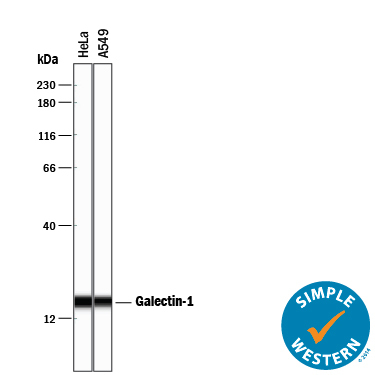
- Detection of Human Galectin-1 by Simple WesternTM. Simple Western lane view shows lysates of HeLa human cervical epithelial carcinoma cell line and A549 human lung carcinoma cell line, loaded at 0.2 mg/mL. A specific band was detected for Galectin-1 at approximately 17 kDa (as indicated) using 1 µg/mL of Goat Anti-Human Galectin-1 Antigen Affinity-purified Polyclonal Antibody (Catalog # AF1152) followed by 1:50 dilution of HRP-conjugated Anti-Goat IgG Secondary Antibody (Catalog # HAF109). This experiment was conducted under reducing conditions and using the 12-230 kDa separation system.
- Submitted by
- Novus Biologicals (provider)
- Main image

- Experimental details
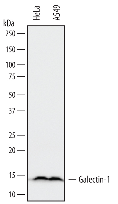
- Detection of Human Galectin-1 by Western Blot. Western blot shows lysates of HeLa human cervical epithelial carcinoma cell line and A549 human lung carcinoma cell line. PVDF membrane was probed with 0.1 µg/mL of Goat Anti-Human Galectin-1 Antigen Affinity-purified Polyclonal Antibody (Catalog # AF1152) followed by HRP-conjugated Anti-Goat IgG Secondary Antibody (Catalog # HAF109). A specific band was detected for Galectin-1 at approximately 14 kDa (as indicated). This experiment was conducted under reducing conditions and using Immunoblot Buffer Group 1.
Supportive validation
- Submitted by
- Novus Biologicals (provider)
- Main image

- Experimental details
- Galectin-1 in Human Prostate Cancer Tissue. Galectin-1 was detected in immersion fixed paraffin-embedded sections of human prostate cancer tissue using 1.7 µg/mL Goat Anti-Human Galectin-1 Antigen Affinity-purified Polyclonal Antibody (Catalog # AF1152) overnight at 4 °C. Tissue was stained with the Anti-Goat HRP-DAB Cell & Tissue Staining Kit (brown; Catalog # CTS008) and counterstained with hematoxylin (blue). View our protocol for Chromogenic IHC Staining of Paraffin-embedded Tissue Sections.

- Submitted by
- Novus Biologicals (provider)
- Main image

- Experimental details
- Galectin-1 in Human Prostate Cancer Tissue. Galectin-1 was detected in immersion fixed paraffin-embedded sections of human prostate cancer tissue using Goat Anti-Human Galectin-1 Antigen Affinity-purified Polyclonal Antibody (Catalog # AF1152) at 5 µg/mL overnight at 4 °C. Tissue was stained using the Anti-Goat HRP-DAB Cell & Tissue Staining Kit (brown; Catalog # CTS008) and counterstained with hematoxylin (blue). Specific labeling was localized to the cytoplasm of stromal cells and nuclei of epithelial cells. View our protocol for Chromogenic IHC Staining of Paraffin-embedded Tissue Sections.

Supportive validation
- Submitted by
- Novus Biologicals (provider)
- Main image

- Experimental details
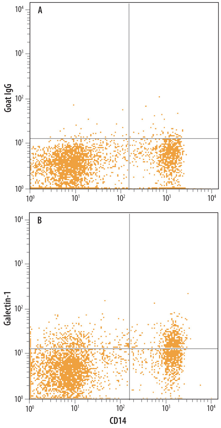
- Detection of Galectin-1 in Human Blood Monocytes by Flow Cytometry. Human peripheral blood monocytes were stained with Mouse Anti-Human CD14 PE-conjugated Monoclonal Antibody (Catalog # FAB3832P) and either (A) Normal Goat IgG Control (Catalog # AB-108-C) or (B) Goat Anti-Human Galectin-1 Antigen Affinity-purified Polyclonal Antibody (Catalog # AF1152) followed by Allophycocyanin-conjugated Anti-Goat IgG Secondary Antibody (Catalog # F0108). To facilitate intracellular staining, cells were fixed with paraformaldehyde and permeabilized with saponin.