 Explore
 Validate
 Learn
Learn Western blot
Antibody data
- Antibody Data
- Antigen structure
- References [7]
- Comments [0]
- Validations
- Western blot [3]
- Immunocytochemistry [2]
- Immunohistochemistry [3]
- Flow cytometry [1]
- Other assay [5]
Submit
Validation data
Reference
Comment
Report error
- Product number
- 43-7400 - Provider product page

- Provider
- Invitrogen Antibodies
- Product name
- Galectin 1 Monoclonal Antibody (6C8.4-1)
- Antibody type
- Monoclonal
- Antigen
- Synthetic peptide
- Reactivity
- Human, Mouse
- Host
- Mouse
- Isotype
- IgG
- Antibody clone number
- 6C8.4-1
- Vial size
- 100 µg
- Concentration
- 0.5 mg/mL
- Storage
- Store at 4°C short term. For long term storage, store at -20°C, avoiding freeze/thaw cycles.
Submitted references Stromal Galectin-1 Promotes Colorectal Cancer Cancer-Initiating Cell Features and Disease Dissemination Through SOX9 and β-Catenin: Development of Niche-Based Biomarkers.
Therapeutic Benefit of Galectin-1: Beyond Membrane Repair, a Multifaceted Approach to LGMD2B.
New Treatment Strategy Targeting Galectin-1 against Thyroid Cancer.
Treatment with galectin-1 improves myogenic potential and membrane repair in dysferlin-deficient models.
Sum of peak intensities outperforms peak area integration in iTRAQ protein expression measurement by LC-MS/MS using a TripleTOF 5600+ platform.
Galectin-1 inhibition attenuates profibrotic signaling in hypoxia-induced pulmonary fibrosis.
Contact-dependent interference with invariant NKT cell activation by herpes simplex virus-infected cells.
Peng KY, Jiang SS, Lee YW, Tsai FY, Chang CC, Chen LT, Yen BL
Frontiers in oncology 2021;11:716055
Frontiers in oncology 2021;11:716055
Therapeutic Benefit of Galectin-1: Beyond Membrane Repair, a Multifaceted Approach to LGMD2B.
Vallecillo-Zúniga ML, Poulson PD, Luddington JS, Arnold CJ, Rathgeber M, Kartchner BC, Hayes S, Gill H, Valdoz JC, Spallino JL, Garfield S, Dodson EL, Arthur CM, Stowell SR, Van Ry PM
Cells 2021 Nov 17;10(11)
Cells 2021 Nov 17;10(11)
New Treatment Strategy Targeting Galectin-1 against Thyroid Cancer.
Gheysen L, Soumoy L, Trelcat A, Verset L, Journe F, Saussez S
Cells 2021 May 5;10(5)
Cells 2021 May 5;10(5)
Treatment with galectin-1 improves myogenic potential and membrane repair in dysferlin-deficient models.
Vallecillo-Zúniga ML, Rathgeber MF, Poulson PD, Hayes S, Luddington JS, Gill HN, Teynor M, Kartchner BC, Valdoz J, Stowell C, Markham AR, Arthur C, Stowell S, Van Ry PM
PloS one 2020;15(9):e0238441
PloS one 2020;15(9):e0238441
Sum of peak intensities outperforms peak area integration in iTRAQ protein expression measurement by LC-MS/MS using a TripleTOF 5600+ platform.
Burat B, Gonzalez J, Sauvage FL, Aouad H, Arnion H, Pinault E, Marquet P, Essig M
Bioscience reports 2019 Jun 28;39(6)
Bioscience reports 2019 Jun 28;39(6)
Galectin-1 inhibition attenuates profibrotic signaling in hypoxia-induced pulmonary fibrosis.
Kathiriya JJ, Nakra N, Nixon J, Patel PS, Vaghasiya V, Alhassani A, Tian Z, Allen-Gipson D, Davé V
Cell death discovery 2017;3:17010
Cell death discovery 2017;3:17010
Contact-dependent interference with invariant NKT cell activation by herpes simplex virus-infected cells.
Bosnjak L, Sahlström P, Paquin-Proulx D, Leeansyah E, Moll M, Sandberg JK
Journal of immunology (Baltimore, Md. : 1950) 2012 Jun 15;188(12):6216-24
Journal of immunology (Baltimore, Md. : 1950) 2012 Jun 15;188(12):6216-24
No comments: Submit comment
Supportive validation
- Submitted by
- Invitrogen Antibodies (provider)
- Main image
- Experimental details
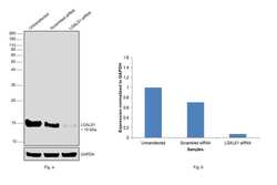
- Knockdown of Galectin 1 was achieved by transfecting PC-3 with Galectin 1 specific siRNAs (Silencer® select Product # s194592, s8145). Western blot analysis (Fig. a) was performed using whole cell extracts (40 ug lysate) from the Galectin 1 knockdown cells (Lane 3), non-targeting scrambled siRNA transfected cells (Lane 2) and untransfected cells (Lane 1). The blot was probed with Galectin 1 Monoclonal Antibody (6C8.4-1) (Product # 43-7400, 1:1000 dilution ) and Goat anti-Mouse IgG (H+L) Superclonal™ Recombinant Secondary Antibody, HRP (Product # A28177, 1:8000 dilution). Densitometric analysis of this western blot is shown in histogram (Fig. b). Decrease in signal upon siRNA mediated knock down confirms that antibody is specific to Galectin 1.
- Submitted by
- Invitrogen Antibodies (provider)
- Main image
- Experimental details
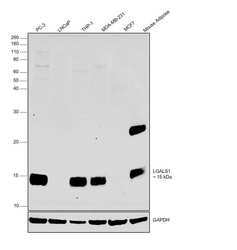
- Western blot was performed using Anti-Galectin 1 Monoclonal Antibody (6C8.4-1) (Product # 43-7400) and a 15kDa band corresponding to Galectin 1 was observed across PC-3, THP-1, MDA-MB-231 but not in LNCaP and MCF7 and in Mouse Adipose. Whole cell extracts (30 µg lysate) of PC-3 (Lane 1), LNCaP (Lane 2), THP-1 (Lane 3), MDA-MB-231 (Lane 4), MCF7 (Lane 5) and tissue extracts (30 ug lysate) of Mouse Adipose (Lane 6) were electrophoresed using NuPAGE™ 12% Bis-Tris Protein Gel (Product # NP0341BOX). Resolved proteins were then transferred onto a Nitrocellulose membrane (Product # IB23001) by iBlot® 2 Dry Blotting System (Product # IB21001). The blot was probed with the primary antibody (1:1000 dilution) and detected by chemiluminescence with Goat anti-Mouse IgG (H+L) Superclonal™ Recombinant Secondary Antibody, HRP (Product # A28177,1:8000 dilution) using the iBright FL 1000 (Product # A32752). Chemiluminescent detection was performed using Novex® ECL Chemiluminescent Substrate Reagent Kit (Product # WP20005). Increased expression of Galectin-1 is seen in triple negative breast cancer (TNCB) (like MDA-MB-231) for promoting metastasis but not in non-TNBC like MCF7 [10.18632/oncotarget.16208] and in androgen unresponsive PC-3 cells in comparison to androgen responsive LNCaP [10.1155/2013/519436]. The band at ~25 kDa in Mouse Adipose corresponds to circulating IgGs that are commonly detected in mouse tissue lysates.
- Submitted by
- Invitrogen Antibodies (provider)
- Main image

- Experimental details
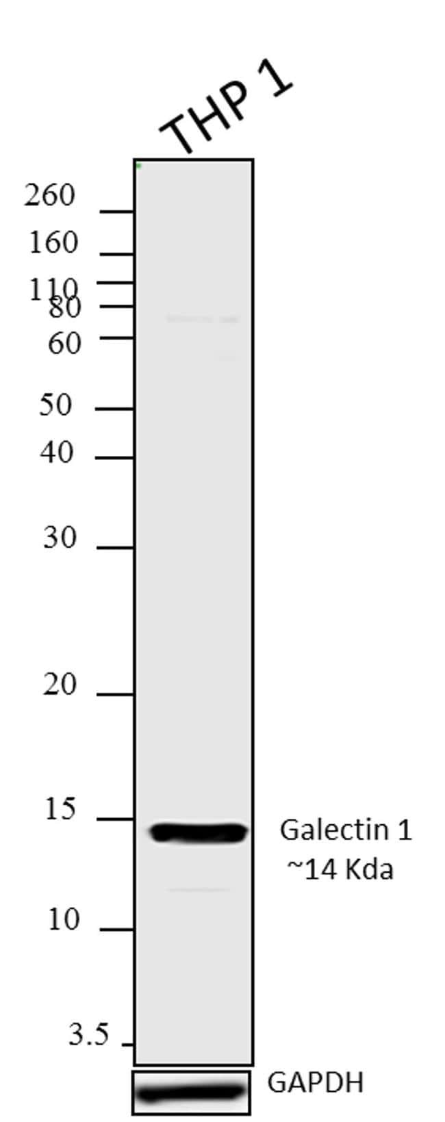
- Western blot analysis was performed on whole cell extracts (30 µg lysate) of THP-1. The blots were probed with Anti-Galectin 1 Mouse Monoclonal Antibody (Product # 43-7400, 1-2 µg/mL) and detected by chemiluminescence Goat anti-Mouse IgG (H+L) Secondary Antibody, HRP conjugate (Product # 62-6520, 1:4000 dilution). A 14 kDa band corresponding to Galectin 1 was observed across cell lines tested.Known quantity of protein samples were electrophoresed using Novex® NuPAGE® 12 % Bis-Tris gel (Product # NP0342BOX), XCell SureLock™ Electrophoresis System (Product # EI0002) and Novex® Sharp Pre-Stained Protein Standard (Product # LC5800). Resolved proteins were then transferred onto a nitrocellulose membrane using iBlot® 2 Dry Blotting System (Product # IB21001). The membrane was probed with the relevant primary and secondary Antibody following blocking with 5 % skimmed milk. Chemiluminescent detection was performed using Pierce™ ECL Western Blotting Substrate (Product # 32106).
Supportive validation
- Submitted by
- Invitrogen Antibodies (provider)
- Main image
- Experimental details
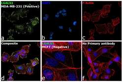
- Immunofluorescence analysis of Galectin 1 was performed using 70% confluent log phase MDA-MB-231 and MCF7 cells. The cells were fixed with 4% paraformaldehyde for 10 minutes, permeabilized with 0.1% Triton™ X-100 for 10 minutes, and blocked with 2% BSA for 45 minutes at room temperature. The cells were labeled with Galectin 1 Monoclonal Antibody (6C8.4-1) (Product # 43-7400) at 1:100 in 0.1% BSA, incubated at 4 degree celsius overnight and then labeled with Donkey anti-Mouse IgG (H+L) Highly Cross-Adsorbed Secondary Antibody, Alexa Fluor Plus 488 (Product # A32766), (1:2500 dilution), for 45 minutes at room temperature (Panel a: Green). Nuclei (Panel b: Blue) were stained with ProLong™ Diamond Antifade Mountant with DAPI (Product # P36962). F-actin (Panel c: Red) was stained with Rhodamine Phalloidin (Product # R415, 1:300). Panel d represents the merged image showing cytoplasmic and membrane localization of Galectin 1 in MDA-MB-231. Panel e represents MCF7 cells showing negative staining for the same. Panel f represents control MDA-MB-231 cells with no primary antibody to assess background. The images were captured at 60X magnification.
- Submitted by
- Invitrogen Antibodies (provider)
- Main image

- Experimental details
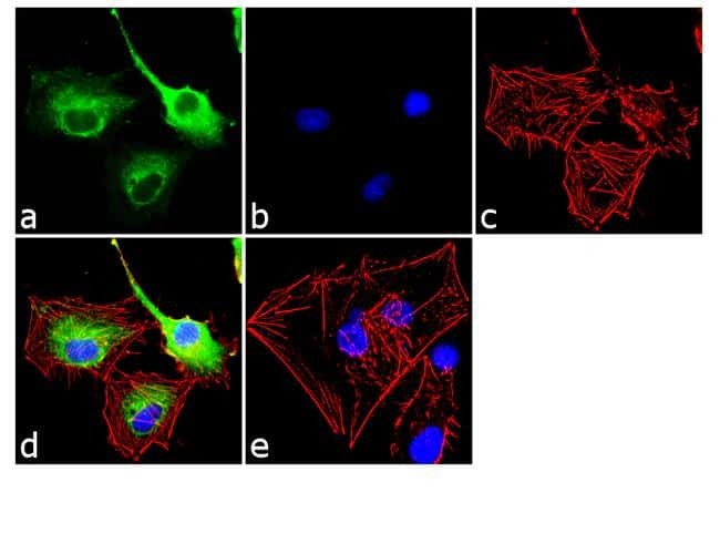
- Immunofluorescence analysis of Galectin 1 was done on 70% confluent log phase U-87MG cells. The cells were fixed with 4% paraformaldehyde for 10 minutes, permeabilized with 0.1% Triton™ X-100 for 10 minutes, and blocked with 1% BSA for 1 hour at room temperature. The cells were labeled with Galectin 1 (6C8.4-1) Mouse Monoclonal Antibody (Product # 43-7400) at 2 µg/mL in 0.1% BSA and incubated for 3 hours at room temperature and then labeled with Goat anti-Mouse IgG (H+L) Superclonal™ Secondary Antibody, Alexa Fluor® 488 conjugate (Product # A28175) at a dilution of 1:2000 for 45 minutes at room temperature (Panel a: green). Nuclei (Panel b: blue) were stained with SlowFade® Gold Antifade Mountant with DAPI (Product # S36938). F-actin (Panel c: red) was stained with Alexa Fluor® 555 Rhodamine Phalloidin (Product # R415, 1:300). Panel d is a merged image showing cytoplasmic localization. Panel e is a no primary antibody control. The images were captured at 60X magnification.
Supportive validation
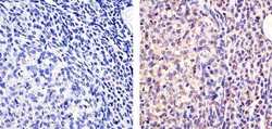
- Submitted by
- Invitrogen Antibodies (provider)
- Main image
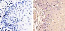
- Experimental details
- Immunohistochemistry analysis of Galectin 1 showing staining in the cytoplasm and nucleus of paraffin-embedded human esophagus tissue (right) compared to a negative control without primary antibody (left). To expose target proteins, antigen retrieval was performed using 10mM sodium citrate (pH 6.0), microwaved for 8-15 min. Following antigen retrieval, tissues were blocked in 3% H2O2-methanol for 15 min at room temperature, washed with ddH2O and PBS, and then probed with a Anti- Galectin 1 Monoclonal Antibody (Product # 43-7400) diluted in 3% BSA-PBS at a dilution of 1:20 overnight at 4°C in a humidified chamber. Tissues were washed extensively in PBST and detection was performed using an HRP-conjugated secondary antibody followed by colorimetric detection using a DAB kit. Tissues were counterstained with hematoxylin and dehydrated with ethanol and xylene to prep for mounting.
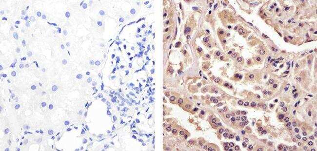
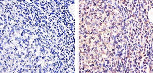
- Submitted by
- Invitrogen Antibodies (provider)
- Main image
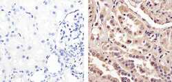
- Experimental details
- Immunohistochemistry analysis of Galectin 1 showing staining in the cytoplasm and nucleus of paraffin-embedded human kidney tissue (right) compared to a negative control without primary antibody (left). To expose target proteins, antigen retrieval was performed using 10mM sodium citrate (pH 6.0), microwaved for 8-15 min. Following antigen retrieval, tissues were blocked in 3% H2O2-methanol for 15 min at room temperature, washed with ddH2O and PBS, and then probed with a Anti- Galectin 1 Monoclonal Antibody (Product # 43-7400) diluted in 3% BSA-PBS at a dilution of 1:100 overnight at 4°C in a humidified chamber. Tissues were washed extensively in PBST and detection was performed using an HRP-conjugated secondary antibody followed by colorimetric detection using a DAB kit. Tissues were counterstained with hematoxylin and dehydrated with ethanol and xylene to prep for mounting.
- Submitted by
- Invitrogen Antibodies (provider)
- Main image
- Experimental details
- Immunohistochemistry analysis of Galectin 1 showing staining in the cytoplasm and nucleus of paraffin-embedded mouse ovary tissue (right) compared to a negative control without primary antibody (left). To expose target proteins, antigen retrieval was performed using 10mM sodium citrate (pH 6.0), microwaved for 8-15 min. Following antigen retrieval, tissues were blocked in 3% H2O2-methanol for 15 min at room temperature, washed with ddH2O and PBS, and then probed with a Anti- Galectin 1 Monoclonal Antibody (Product # 43-7400) diluted in 3% BSA-PBS at a dilution of 1:50 overnight at 4°C in a humidified chamber. Tissues were washed extensively in PBST and detection was performed using an HRP-conjugated secondary antibody followed by colorimetric detection using a DAB kit. Tissues were counterstained with hematoxylin and dehydrated with ethanol and xylene to prep for mounting.
Supportive validation
- Submitted by
- Invitrogen Antibodies (provider)
- Main image
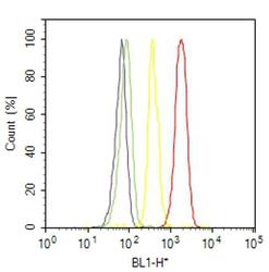
- Experimental details
- Flow cytometry analysis of Galectin 1 was done on U-87MG cells. Cells were fixed with 70% ethanol for 10 minutes, permeabilized with 0.25% Triton™ X-100 for 20 minutes, and blocked with 5% BSA for 30 minutes at room temperature. Cells were labeled with Galectin 1 Mouse Monoclonal Antibody (437400, red histogram) or with mouse isotype control (yellow histogram) at 3-5 ug/million cells in 2.5% BSA. After incubation at room temperature for 2 hours, the cells were labeled with Alexa Fluor® 488 Rabbit Anti-Mouse Secondary Antibody (A11059) at a dilution of 1:400 for 30 minutes at room temperature. The representative 10,000 cells were acquired and analyzed for each sample using an Attune® Acoustic Focusing Cytometer. The purple histogram represents unstained control cells and the green histogram represents no-primary-antibody control.
Supportive validation
- Submitted by
- Invitrogen Antibodies (provider)
- Main image
- Experimental details
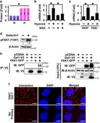
- Figure 5 Galectin-1 and FAK1 mutually activated each other in hypoxic lung epithelial cells. ( a ) Galectin-1 protein levels were increased following hypoxia treatment (1% O 2 ; 72 h) as determined by LC-MS/MS in H441 cells. * P

- Submitted by
- Invitrogen Antibodies (provider)
- Main image
- Experimental details
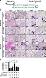
- Figure 6 Hypoxia causes exacerbation of fibrosis via galectin-1 in mice injured with low levels of bleomycin. ( A ) Experimental design depicting two bleomycin insults (day 0--D0; day 7--D7) followed by hypoxia treatment for 3 weeks (day 8 to day 28). Galectin-1 inhibitor (OTX008) treatment was performed from day 5 to day 28. ( B ) Hypoxia treatment of mice injured with bleomycin induced fibrosis. Mice were exposed to hypoxia alone (m-p) or treated with intratracheal bleomycin alone (e-h) or underwent hypoxia and bleomycin insults together (q-t). Mice were also treated with galectin-1 inhibitor (i-l, u-x) or saline (e-h, q-t). H&E staining (a, e, i, m, q, u), Masson's trichrome staining for visualization of collagen deposition (b, f, j, n, r, v), alpha -SMA staining (c, g, k, o, s, w), and galectin-1 staining (d, h, l, p, t, x) was performed. Black scale bars=100 mu m; blue scale bars=20 mu m. alpha -SMA expression is increased in the proximal parenchyma (s; arrows) and distal parenchyma (s, inset) of mouse lungs treated with hypoxia and bleomycin. Similarly, galectin-1 expression is increased in proximal parenchyma (t; arrow and right bottom inset) and distal parenchyma (left bottom inset). Galectin-1 inhibition reversed lung remodeling (u), collagen deposition (v), and reduced alpha -SMA (w) and galectin-1 expression (x). ( C ) Galectin-1 inhibition reduced total collagen deposition in the lungs of mice injured with hypoxia and bleomycin. Total collagen was measured us
- Submitted by
- Invitrogen Antibodies (provider)
- Main image
- Experimental details
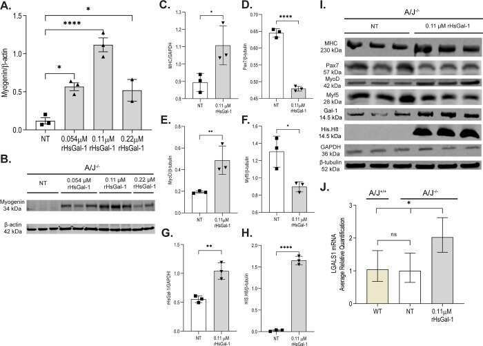
- 10.1371/journal.pone.0238441.g001 Fig 1 rHsGal-1 increases myogenic regulatory factors in A/J -/- myotubes. A. Quantification of myogenin after 72h treatment with varying concentrations of rHsGal-1. B. Western blot images of myogenin at different rHsGal-1 treatments. C-F. Quantification of myogenic markers MHC(C), Pax7(D), MyoD(E), and Myf5(F) in A/J -/- myotubes after 72h treatment with 0.11muM rHsGal-1. G-H. Quantification of Gal-1(G) and His.H8(H)in A/J -/- myotubes after 72h treatment with 0.11muM rHsGal-1. I. Western blot images of myogenic markers (Pax7, Myf5, MyoD, and MHC) and of mouse Gal-1 and His Tagged rHsGal-1. J. RT-qPCR quantification of LGALS1 transcript between A/J WT, A/J -/- NT, and A/J -/- 0.11muM rHsGal-1 treated myotubes. p values are measured by Tukey's multiple comparison test and indicated by *p< 0.05, **p< 0.01, ****p< 0.0001 (n = 3 for each group). Error bars represent SEM.
- Submitted by
- Invitrogen Antibodies (provider)
- Main image
- Experimental details
- NULL
- Submitted by
- Invitrogen Antibodies (provider)
- Main image
- Experimental details
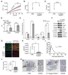
- Figure 4 rHsGal-1 improves membrane repair and exploratory activity and decreases inflammatory markers in Bla/J mice after one-month treatment. ( A ) Quantification of laser injury on muscle from BLA/J mice treated for one month with either 2.7 mg/kg rHsGal-1 or PBS. ( B ) Average number of rearing events during first hour placed in CLAMS cages for 2.7 mg/kg rHsGal-1 treated, and PBS treated BLA/J mice. ( C ) Average number of times the mice crossed the x-axis during first hour placed in CLAMS cages for 2.7 mg/kg rHsGal-1 treated and PBS treated control BLA/J mice. ( D ) RT-qPCR results for Gal-1 gene expression in the psoas of Bla/J mice treated for 1 week or 1 month with PBS (control) or 2.7 mg/kg rHsGal-1. ( E ) Quantification of Western blot comparing levels of Gal-1, His-Tag, p65, P-p65, p50, all normalized to beta-tubulin control. Tissue was taken from Bla/J mice treated with 2.7 mg/kg rHsGal-1 or PBS (control) for one month. ( F ) Western blot images from homogenized muscle tissue from PBS or 2.7 mg/kg rHsGal-1 treated BLA/J mice. ( G ) Representative images of immunofluorescence on Bla/J mice treated for one month with 2.7 mg/kg rHsGal-1 or PBS (control). Samples were stained with p65, DAPI, and Phalloidin. ( H ) Quantification of immunofluorescence of p65 normalized to DAPI control on mouse psoas muscles either treated with 2.7 mg/kg rHsGal-1 or PBS. ( I ) Concentration of Gal-1 present in serum of Bla/J mice treated with rHsGal-1 from time of treatment (t = 0) to 12
