Explore
 Validate
 Learn
Learn Western blot
Antibody data
- Antibody Data
- Antigen structure
- References [3]
- Comments [0]
- Validations
- Western blot [2]
- ELISA [2]
- Immunocytochemistry [1]
- Immunoprecipitation [1]
Submit
Validation data
Reference
Comment
Report error
- Product number
- H00003956-M01 - Provider product page

- Provider
- Novus Biologicals
- Proper citation
- Novus Cat#H00003956-M01, RRID:AB_539039
- Product name
- Mouse Monoclonal Galectin-1 Antibody
- Antibody type
- Monoclonal
- Description
- IgG purified. LGALS1 - lectin, galactoside-binding, soluble, 1 (galectin 1)
- Reactivity
- Human
- Host
- Mouse
- Isotype
- IgG
- Vial size
- 0.1 mg
- Storage
- Aliquot and store at -20C or -80C. Avoid freeze-thaw cycles.
Submitted references Retrospective Proteomic Screening of 100 Breast Cancer Tissues.
Galectin-1 and Galectin-3 Mediate Protocadherin-24-Dependent Membrane Localization of β-catenin in Colon Cancer Cell Line HCT116.
Quantitative and temporal proteome analysis of butyrate-treated colorectal cancer cells.
Pucci-Minafra I, Di Cara G, Musso R, Cancemi P, Albanese NN, Roz E, Minafra S
Proteomes 2017 Jul 7;5(3)
Proteomes 2017 Jul 7;5(3)
Galectin-1 and Galectin-3 Mediate Protocadherin-24-Dependent Membrane Localization of β-catenin in Colon Cancer Cell Line HCT116.
Ose R, Oharaa O, Nagase T
Current chemical genomics 2012;6:18-26
Current chemical genomics 2012;6:18-26
Quantitative and temporal proteome analysis of butyrate-treated colorectal cancer cells.
Tan HT, Tan S, Lin Q, Lim TK, Hew CL, Chung MC
Molecular & cellular proteomics : MCP 2008 Jun;7(6):1174-85
Molecular & cellular proteomics : MCP 2008 Jun;7(6):1174-85
No comments: Submit comment
Supportive validation
- Submitted by
- Novus Biologicals (provider)
- Main image
- Experimental details
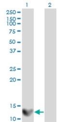
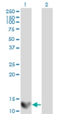
- Western Blot: Galectin-1 Antibody (1E8-1B2) [H00003956-M01] - Analysis of LGALS1 expression in transfected 293T cell line by LGALS1 monoclonal antibody (M01), clone 1E8-1B2.Lane 1: LGALS1 transfected lysate(14.7 KDa).Lane 2: Non-transfected lysate.
- Submitted by
- Novus Biologicals (provider)
- Main image
- Experimental details
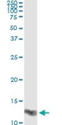
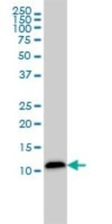
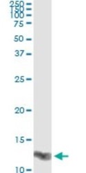
- Western Blot: Galectin-1 Antibody (1E8-1B2) [H00003956-M01] - LGALS1 monoclonal antibody (M01), clone 1E8-1B2 Analysis of LGALS1 expression in HepG2.

Supportive validation
- Submitted by
- Novus Biologicals (provider)
- Main image
- Experimental details
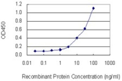
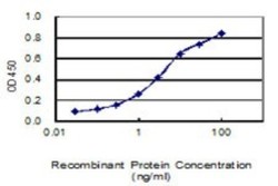
- Sandwich ELISA: Galectin-1 Antibody (1E8-1B2) [H00003956-M01] - Detection limit for recombinant GST tagged LGALS1 is 1 ng/ml as a capture antibody.
- Submitted by
- Novus Biologicals (provider)
- Main image
- Experimental details
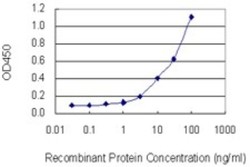
- Sandwich ELISA: Galectin-1 Antibody (1E8-1B2) [H00003956-M01] - Detection limit for recombinant GST tagged LGALS1 is 0.03 ng/ml as a capture antibody.

Supportive validation
- Submitted by
- Novus Biologicals (provider)
- Main image
- Experimental details
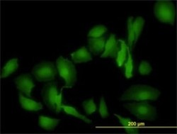
- Immunocytochemistry/Immunofluorescence: Galectin-1 Antibody (1E8-1B2) [H00003956-M01] - Analysis of monoclonal antibody to LGALS1 on HeLa cell. Antibody concentration 10 ug/ml.

Supportive validation
- Submitted by
- Novus Biologicals (provider)
- Main image
- Experimental details
- Immunoprecipitation: Galectin-1 Antibody (1E8-1B2) [H00003956-M01] - Analysis of LGALS1 transfected lysate using anti-LGALS1 monoclonal antibody and Protein A Magnetic Bead, and immunoblotted with LGALS1 MaxPab rabbit polyclonal antibody.