Explore
 Validate
 Learn
Learn Western blot
Antibody data
- Antibody Data
- Antigen structure
- References [0]
- Comments [0]
- Validations
- Western blot [3]
Submit
Validation data
Reference
Comment
Report error
- Product number
- PA5-19665 - Provider product page

- Provider
- Invitrogen Antibodies
- Product name
- Anti-Galectin 1 Polyclonal Antibody
- Antibody type
- Polyclonal
- Antigen
- Synthetic peptide
- Description
- This antibody is predicted to react with mouse, rat, sheep, cow, pig and orangutan based on sequence homology. Store antibody at 4ºC for 1-2 weeks. For long-term storage, store at -20ºC.
- Reactivity
- Human, Hamster
- Host
- Rabbit
- Isotype
- IgG
- Vial size
- 100 µg
- Concentration
- 0.7 mg/mL
- Storage
- Store at 4°C short term. For long term storage, store at -20°C, avoiding freeze/thaw cycles.
No comments: Submit comment
Supportive validation
- Submitted by
- Invitrogen Antibodies (provider)
- Main image
- Experimental details
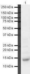
- Western blot analysis of Human Heart Tissue Lysate using Product # PA5-19665, Galectin 1 primary antibody at a dilution of 1 µg/mL. Blot treated with a secondary HRP-conjugated Goat polyclonal anti-Rabbit antibody was used at a dilution of 1:3000.
- Submitted by
- Invitrogen Antibodies (provider)
- Main image
- Experimental details
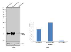
- Knockdown of Galectin 1 (LGALS1) was achieved by transfecting PC-3 with LGALS1 specific siRNAs (Silencer® select Product # s194592, s8145). Western blot analysis (Fig. a) was performed using whole cell extracts from the Galectin 1 knockdown cells (lane 3), non-targeting scrambled siRNA transfected cells (lane 2) and untransfected cells (lane 1). The blot was probed with Galectin 1 Polyclonal Antibody (Product # PA5-19665, 0.7 µg/mL dilution) and Goat anti-Rabbit IgG (H+L) Superclonal™ Recombinant Secondary Antibody, HRP (Product # A27036, 1:10000 dilution). Densitometric analysis of this western blot is shown in histogram (Fig. b). Decrease in signal upon siRNA mediated knock down confirms that antibody is specific to Galectin 1.

- Submitted by
- Invitrogen Antibodies (provider)
- Main image
- Experimental details
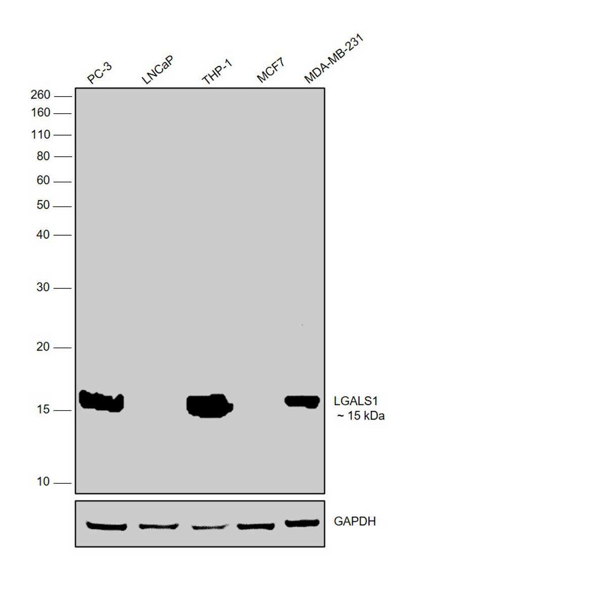
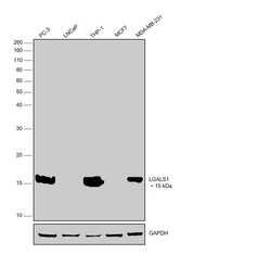
- Western blot was performed using Anti-Galectin 1 Polyclonal Antibody (Product # PA5-19665) and a 15 kDa band corresponding to Galectin 1 was observed across cell lines tested except in MCF7 and LNCaP which are reported low expressing for the same. Whole cell extracts (30 µg lysate) of PC-3 (Lane 1), LNCaP (Lane 2), THP-1 (Lane 3), MCF7 (Lane 4) and MDA-MB-231 (Lane 5) were electrophoresed using NuPAGE™ 12% Bis-Tris Protein Gel (Product # NP0341BOX). Resolved proteins were then transferred onto a Nitrocellulose membrane (Product # IB23001) by iBlot® 2 Dry Blotting System (Product # IB21001). The blot was probed with the primary antibody (0.7 µg/mL dilution) and detected by chemiluminescence with Goat anti-Rabbit IgG (H+L) Superclonal™ Recombinant Secondary Antibody, HRP (Product # A27036, 1:10000 dilution) using the iBright FL 1000 (Product # A32752). Chemiluminescent detection was performed using SuperSignal™ West Dura Extended Duration Substrate (Product # 34076).