 Explore
 Validate
 Learn
Learn Western blot
 Immunocytochemistry
Antibody data
- Antibody Data
- Antigen structure
- References [1]
- Comments [0]
- Validations
- Immunocytochemistry [6]
- Immunoprecipitation [1]
- Other assay [1]
Submit
Validation data
Reference
Comment
Report error
- Product number
- PA5-30706 - Provider product page

- Provider
- Invitrogen Antibodies
- Product name
- Galectin 1 Polyclonal Antibody
- Antibody type
- Polyclonal
- Antigen
- Recombinant full-length protein
- Description
- Recommended positive controls: K562, THP-1, HL-60, 293T, NCI-H929, BCL-1, PC-12, Rat2. Predicted reactivity: Mouse (88%), Rat (90%), Dog (82%), Pig (85%), Sheep (85%), Rhesus Monkey (97%), Bovine (86%). Store product as a concentrated solution. Centrifuge briefly prior to opening the vial.
- Reactivity
- Human, Mouse, Rat
- Host
- Rabbit
- Isotype
- IgG
- Vial size
- 100 μL
- Concentration
- 1.48 mg/mL
- Storage
- Store at 4°C short term. For long term storage, store at -20°C, avoiding freeze/thaw cycles.
Submitted references Quantitative proteomic analysis of the brainstem following lethal sarin exposure.
Meade ML, Hoffmann A, Makley MK, Snider TH, Schlager JJ, Gearhart JM
Brain research 2015 Jun 22;1611:101-13
Brain research 2015 Jun 22;1611:101-13
No comments: Submit comment
Supportive validation
- Submitted by
- Invitrogen Antibodies (provider)
- Main image
- Experimental details
- Immunofluorescent analysis of Galectin 1 in methanol-fixed A431 cells using a Galectin 1 polyclonal antibody (Product # PA5-30706) at a 1:200 dilution.

- Submitted by
- Invitrogen Antibodies (provider)
- Main image
- Experimental details
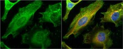
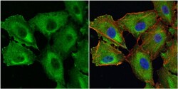
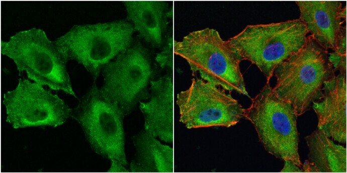
- Immunocytochemistry-Immunofluorescence analysis of Galectin 1 was performed in HeLa cells fixed in 4% paraformaldehyde at RT for 15 min. Green: Galectin 1 Polyclonal Antibody (Product # PA5 30706) diluted at 1:200. Red: phalloidin, a cytoskeleton marker. Blue: Hoechst 33342 staining.

- Submitted by
- Invitrogen Antibodies (provider)
- Main image

- Experimental details
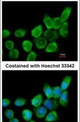
- Immunocytochemistry-Immunofluorescence analysis of Galectin 1 was performed in A549 cells fixed in 4% paraformaldehyde at RT for 15 min. Green: Galectin 1 Polyclonal Antibody (Product # PA5 30706) diluted at 1:500. Red: phalloidin, a cytoskeleton marker. Blue: Hoechst 33342 staining.

- Submitted by
- Invitrogen Antibodies (provider)
- Main image
- Experimental details
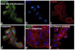
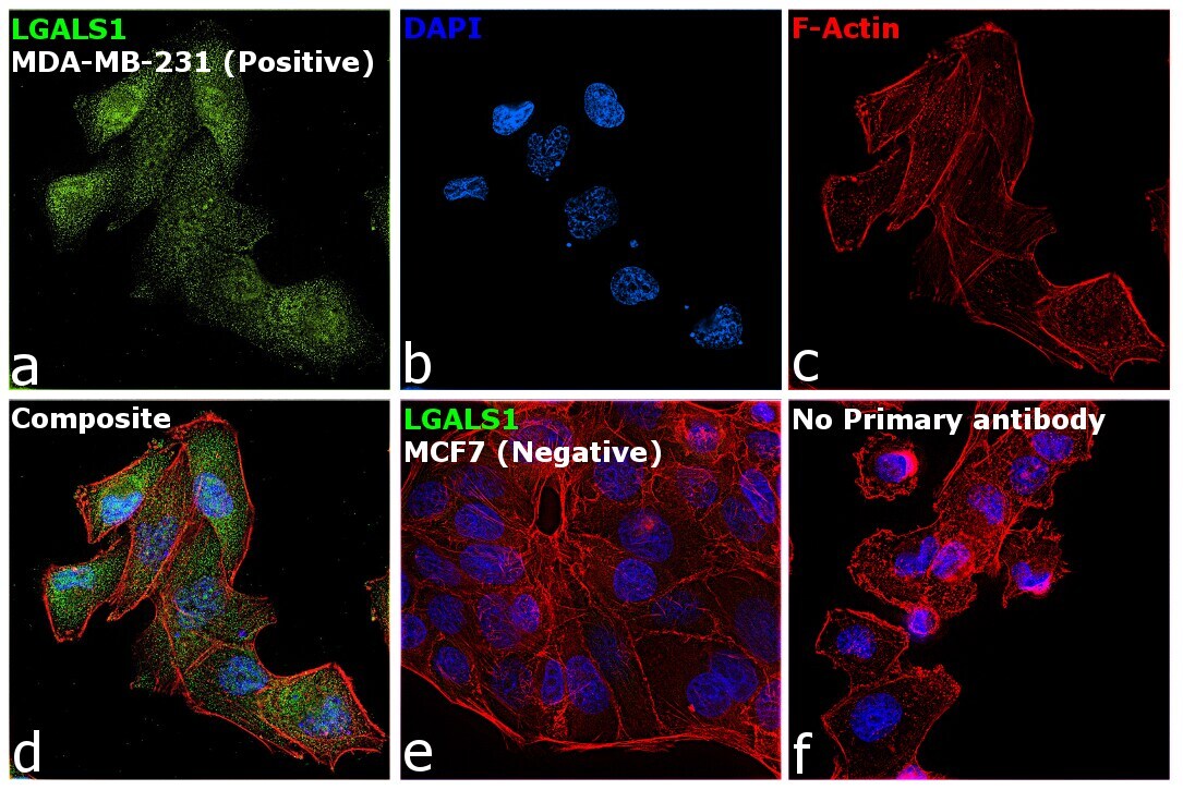
- Immunofluorescence analysis of Galectin 1 was performed using 70% confluent log phase MDA-MB-231 and MCF7 cells. The cells were fixed with 4% paraformaldehyde for 10 minutes, permeabilized with 0.1% Triton™ X-100 for 10 minutes, and blocked with 2% BSA for 45 minutes at room temperature. The cells were labeled with Galectin 1 Polyclonal Antibody (Product # PA5-30706) at 1:200 in 0.1% BSA, incubated at 4 degree celsius overnight and then labeled with Donkey anti-Rabbit IgG (H+L) Highly Cross-Adsorbed Secondary Antibody, Alexa Fluor Plus 488 (Product # A32790), (1:2500 dilution), for 45 minutes at room temperature (Panel a: Green). Nuclei (Panel b: Blue) were stained with ProLong™ Diamond Antifade Mountant with DAPI (Product # P36962). F-actin (Panel c: Red) was stained with Rhodamine Phalloidin (Product # R415, 1:300). Panel d represents the merged image showing cytoplasmic localization of Galectin 1 in MDA-MB-231. Panel e represents MCF7 cells showing negative staining for the same. Panel f represents control MDA-MB-231 cells with no primary antibody to assess background. The images were captured at 60X magnification.

- Submitted by
- Invitrogen Antibodies (provider)
- Main image
- Experimental details
- Immunocytochemistry-Immunofluorescence analysis of Galectin 1 was performed in A549 cells fixed in 4% paraformaldehyde at RT for 15 min. Green: Galectin 1 Polyclonal Antibody (Product # PA5 30706) diluted at 1:500. Red: phalloidin, a cytoskeleton marker. Blue: Hoechst 33342 staining.
- Submitted by
- Invitrogen Antibodies (provider)
- Main image

- Experimental details
- Immunofluorescence analysis of Galectin 1 was performed using 70% confluent log phase MDA-MB-231 and MCF7 cells. The cells were fixed with 4% paraformaldehyde for 10 minutes, permeabilized with 0.1% Triton™ X-100 for 10 minutes, and blocked with 2% BSA for 45 minutes at room temperature. The cells were labeled with Galectin 1 Polyclonal Antibody (Product # PA5-30706) at 1:200 in 0.1% BSA, incubated at 4 degree celsius overnight and then labeled with Donkey anti-Rabbit IgG (H+L) Highly Cross-Adsorbed Secondary Antibody, Alexa Fluor Plus 488 (Product # A32790), (1:2500 dilution), for 45 minutes at room temperature (Panel a: Green). Nuclei (Panel b: Blue) were stained with ProLong™ Diamond Antifade Mountant with DAPI (Product # P36962). F-actin (Panel c: Red) was stained with Rhodamine Phalloidin (Product # R415, 1:300). Panel d represents the merged image showing cytoplasmic localization of Galectin 1 in MDA-MB-231. Panel e represents MCF7 cells showing negative staining for the same. Panel f represents control MDA-MB-231 cells with no primary antibody to assess background. The images were captured at 60X magnification.
Supportive validation
- Submitted by
- Invitrogen Antibodies (provider)
- Main image

- Experimental details
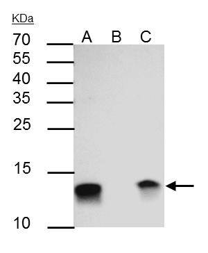
- Galectin 1 Polyclonal Antibody immunoprecipitates Galectin 1 protein in IP experiments. IP Sample: HeLa whole cell lysate/extract. A : 30 µg whole cell lysate/extract of Galectin 1 Polyclonal Antibody expressing HeLa cells. B : Control with 2.5 µg of pre-immune rabbit IgG. C : Immunoprecipitation of Galectin 1 by 2.5 µg of Galectin 1 Polyclonal Antibody (Product # PA5-30706). 15% SDS-PAGE. The immunoprecipitated Galectin 1 protein was detected by Galectin 1 Polyclonal Antibody (Product # PA5-30706) diluted at 1:1,000. Anti-rabbit IgG (HRP) was used as a secondary reagent.
Supportive validation
- Submitted by
- Invitrogen Antibodies (provider)
- Main image

- Experimental details
- Galectin 1 Polyclonal Antibody immunoprecipitates Galectin 1 protein in IP experiments. IP Sample: HeLa whole cell lysate/extract. A : 30 µg whole cell lysate/extract of Galectin 1 Polyclonal Antibody expressing HeLa cells. B : Control with 2.5 µg of pre-immune rabbit IgG. C : Immunoprecipitation of Galectin 1 by 2.5 µg of Galectin 1 Polyclonal Antibody (Product # PA5-30706). 15% SDS-PAGE. The immunoprecipitated Galectin 1 protein was detected by Galectin 1 Polyclonal Antibody (Product # PA5-30706) diluted at 1:1,000. Anti-rabbit IgG (HRP) was used as a secondary reagent.
