Explore
 Validate
 Learn
Learn Western blot
 Immunocytochemistry
Antibody data
- Antibody Data
- Antigen structure
- References [0]
- Comments [0]
- Validations
- Western blot [1]
Submit
Validation data
Reference
Comment
Report error
- Product number
- PA1422 - Provider product page

- Provider
- Boster Biological Technology
- Product name
- Anti-Galectin 1/LGALS1 Antibody
- Antibody type
- Polyclonal
- Description
- Polyclonal antibody for GALECTIN 1/LGALS1 detection. Host: Rabbit.Size: 100μg/vial. Tested applications: WB, IHC-P, IHC-F, ICC/IF, FCM. Reactive species: Human. GALECTIN 1/LGALS1 information: Molecular Weight: 14716 MW; Subcellular Localization: Secreted, extracellular space, extracellular matrix ; Tissue Specificity: Expressed in placenta, maternal decidua and fetal membranes. Within placenta, expressed in trophoblasts, stromal cells, villous endothelium, syncytiotrophoblast apical membrane and villous stroma. Within fetal membranes, expressed in amnion, chorioamniotic mesenchyma and chorion (at protein level). Expressed in cardiac, smooth, and skeletal muscle, neurons, thymus, kidney and hematopoietic cells.
- Reactivity
- Human
- Host
- Rabbit
- Vial size
- 100μg/vial
- Concentration
- Add 0.2ml of distilled water will yield a concentration of 500ug/ml.
- Storage
- At -20°C for one year. After reconstitution, at 4°C for one month. It can also be aliquoted and stored frozen at -20°C for a longer time. Avoid repeated freezing and thawing.
- Handling
- Add 0.2ml of distilled water will yield a concentration of 500ug/ml.
No comments: Submit comment
Supportive validation
- Submitted by
- Boster Biological Technology (provider)
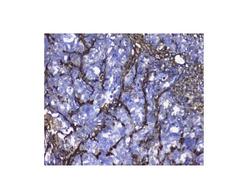
- Main image
- Experimental details
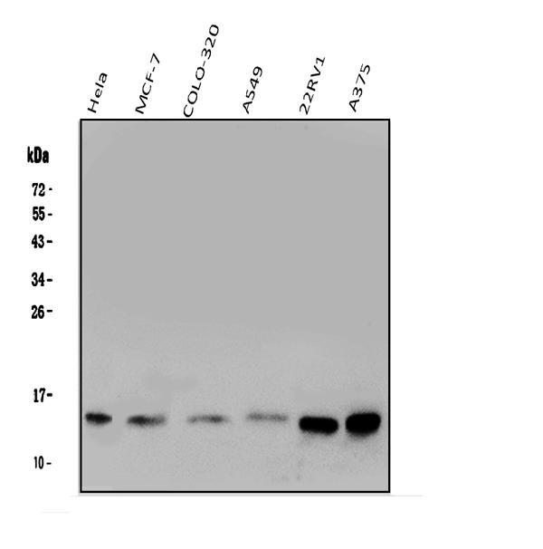
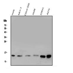
- Western blot analysis of LGALS1 using anti-LGALS1 antibody (PA1422). Electrophoresis was performed on a 5-20% SDS-PAGE gel at 70V (Stacking gel) / 90V (Resolving gel) for 2-3 hours. The sample well of each lane was loaded with 50ug of sample under reducing conditions. Lane 1: human Hela whole cell lysates, Lane 2: human MCF-7 whole cell lysates, Lane 3: human COLO-320 whole cell lysates, Lane 4: human A549 whole cell lysates, Lane 5: human 22RV1 whole cell lysates; Lane 6: human A375 whole cell lysates. After Electrophoresis, proteins were transferred to a Nitrocellulose membrane at 150mA for 50-90 minutes. Blocked the membrane with 5% Non-fat Milk/ TBS for 1.5 hour at RT. The membrane was incubated with rabbit anti-LGALS1 antigen affinity purified polyclonal antibody (Catalog # PA1422) at 0.5 μg/mL overnight at 4°C, then washed with TBS-0.1%Tween 3 times with 5 minutes each and probed with a goat anti-rabbit IgG-HRP secondary antibody at a dilution of 1:10000 for 1.5 hour at RT. The signal is developed using an Enhanced Chemiluminescent detection (ECL) kit (Catalog # EK1002) with Tanon 5200 system. A specific band was detected for LGALS1 at approximately 15KD. The expected band size for LGALS1 is at 15KD.
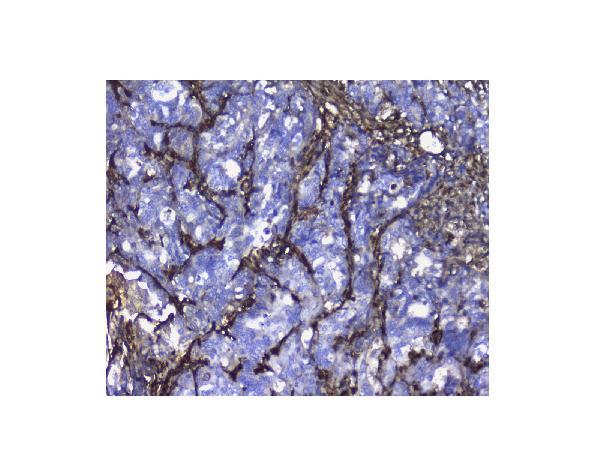
- Additional image