Explore
 Validate
 Learn
Learn Western blot
 ELISA
ELISA Immunocytochemistry
Antibody data
- Antibody Data
- Antigen structure
- References [1]
- Comments [0]
- Validations
- Immunocytochemistry [1]
- Other assay [1]
Submit
Validation data
Reference
Comment
Report error
- Product number
- PA5-96231 - Provider product page

- Provider
- Invitrogen Antibodies
- Product name
- VGAT Polyclonal Antibody
- Antibody type
- Polyclonal
- Antigen
- Recombinant full-length protein
- Description
- Positive Samples: Mouse spinal cord, Mouse brain; Cellular Location: Cytoplasmic vesicle membrane, Multi-pass membrane protein Immunogen sequence: MATLLRSKLS NVATSVSNKS QAKMSGMFAR MGFQAATDEE AVGFAHCDDL DFEHRQGLQM DILKAEGEPC GDEGAEAPVE GDIHYQRGSG APLPPSGSKD QVGGGGEFGG HDKPKITAWE AGWNVTNAIQ GMFVLGLPYA ILHGGYLGLF LIIFAAVVCC YTGKILIACL YEENEDGEVV RVRDSYVAIA NACCAPRFPT LGGRVVNVAQ IIELVMTCIL YVVVSGNLMY NSFPGLPVSQ KSWSIIATAV LLPCAFLKNL KAVSKFSLLC TLAHFVINIL VIAYCLSRAR DWAWEKVKFY IDVKKFPISI GIIVFSYTSQ IFLPSLEGNM QQPSEFHCMM NWTHIAACVL KGLFALVAYL TWADETKEVI TDNLPGSIRA VVNIFLVAKA LLSYPLPFFA AVEVLEKSLF QEGSRAFFPA CYSGDGRLKS WGLTLRCALV VFTLLMAIYV PHFALLMGLT GSLTGAGLCF LLPSLFHLRL LWRKLLWHQV FFDVAIFVIG GICSVSGFVH SLEGLIEAYR TNAED
- Reactivity
- Human, Mouse, Rat
- Host
- Rabbit
- Isotype
- IgG
- Vial size
- 100 μL
- Concentration
- 2.79 mg/mL
- Storage
- -20°C, Avoid Freeze/Thaw Cycles
Submitted references Restoring tripartite glutamatergic synapses: A potential therapy for mood and cognitive deficits in Gulf War illness.
Wang X, Xu Z, Zhao F, Lin KJ, Foster JB, Xiao T, Kung N, Askwith CC, Bruno JP, Valentini V, Hodgetts KJ, Lin CG
Neurobiology of stress 2020 Nov;13:100240
Neurobiology of stress 2020 Nov;13:100240
No comments: Submit comment
Supportive validation
- Submitted by
- Invitrogen Antibodies (provider)
- Main image
- Experimental details
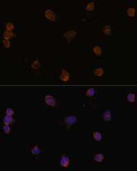
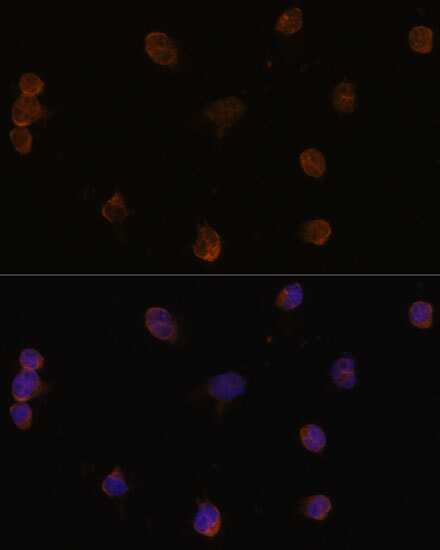
- Immunocytochemistry-Immunofluorescence analysis of VGAT was performed in Y79 cells using VGAT Polyclonal Antibody (Product # PA5-96231) at a dilution of 1:100. Blue: DAPI for nuclear staining.
Supportive validation
- Submitted by
- Invitrogen Antibodies (provider)
- Main image

- Experimental details
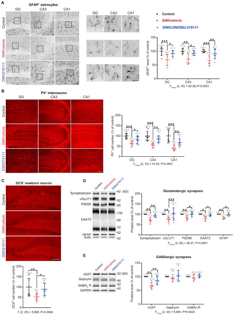
- Fig. 7 GW-exposure causes impaired glutamatergic synapses, astrocyte atrophy, loss of interneurons, and decreased neurogenesis, which can be restored by LDN/OSU-215111. Upon completion of the behavioral assessments in Fig. 5 , a subset of mice was euthanized for the following pathological studies. (A) GFAP immunohistochemical staining to examine astrocytes. Representative images are shown (scale bars: 100 mum). Boxed areas are shown enlarged in the middle column (scale bars: 25 mum). Quantitative analysis indicates that the GFAP + area decreased in GWI/vehicle mice and was partially restored in LDN/OSU-215111 treated mice. Data was generated from the percentage of GFAP staining in 890 mum x 667 mum area and normalized by control. n = 5-6 animals per group; average of >=6 sections per animal. (B) Parvalbumin immunohistochemical staining to examine parvalbumin-expressing interneurons. Representative images are shown (scale bars: 100 mum). Quantitative analysis indicates that the number of PV + interneurons decreased in GWI/vehicle mice and was partially restored in LDN/OSU-215111 treated mice. n = 9-10 animals per group; average of >=6 sections per animal. (C) Doublecortin immunohistochemical staining to examine newborn neurons in the dentate gyrus. Representative images are shown (scale bars: 100 mum). Quantitative analysis indicates that the number of DCX + cells decreased in GWI/vehicle mice and was partially restored in LDN/OSU-215111 treated mice. n = 9-10 animals per grou