Explore
 Validate
 Learn
Learn Western blot
 Immunocytochemistry
Antibody data
- Antibody Data
- Antigen structure
- References [2]
- Comments [0]
- Validations
- Immunocytochemistry [4]
- Immunohistochemistry [5]
- Other assay [1]
Submit
Validation data
Reference
Comment
Report error
- Product number
- PA5-27569 - Provider product page

- Provider
- Invitrogen Antibodies
- Product name
- VGAT Polyclonal Antibody
- Antibody type
- Polyclonal
- Antigen
- Recombinant full-length protein
- Description
- Recommended positive controls: SLC32A1-transfected 293T. Predicted reactivity: Mouse (97%), Rat (95%), Xenopus laevis (83%), Rhesus Monkey (97%). Store product as a concentrated solution. Centrifuge briefly prior to opening the vial.
- Reactivity
- Human, Mouse, Rat
- Host
- Rabbit
- Isotype
- IgG
- Vial size
- 100 μL
- Concentration
- 0.52 mg/mL
- Storage
- Store at 4°C short term. For long term storage, store at -20°C, avoiding freeze/thaw cycles.
Submitted references Precise Coordination of Three-Dimensional Rotational Kinematics by Ventral Tegmental Area GABAergic Neurons.
Dose-dependent reversal of KCC2 hypofunction and phenobarbital-resistant neonatal seizures by ANA12.
Hughes RN, Watson GDR, Petter EA, Kim N, Bakhurin KI, Yin HH
Current biology : CB 2019 Oct 7;29(19):3244-3255.e4
Current biology : CB 2019 Oct 7;29(19):3244-3255.e4
Dose-dependent reversal of KCC2 hypofunction and phenobarbital-resistant neonatal seizures by ANA12.
Carter BM, Sullivan BJ, Landers JR, Kadam SD
Scientific reports 2018 Aug 10;8(1):11987
Scientific reports 2018 Aug 10;8(1):11987
No comments: Submit comment
Supportive validation
- Submitted by
- Invitrogen Antibodies (provider)
- Main image

- Experimental details
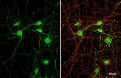
- Immunocytochemistry-Immunofluorescence analysis of VGAT was performed in Cultured rat E18 primary cortical neuron, DIV 8. Cells fixed in 4% paraformaldehyde at RT for 15 min. Green: VGAT Polyclonal Antibody (Product # PA5-27569) diluted at 1:250. Red: beta Tubulin 3/ TUJ1, stained by beta Tubulin 3/ TUJ1 antibody. Blue: Fluoroshield with DAPI.
- Submitted by
- Invitrogen Antibodies (provider)
- Main image
- Experimental details
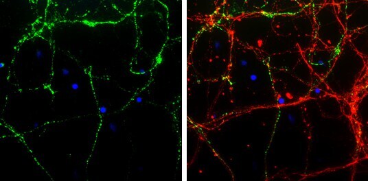
- VGAT Polyclonal Antibody detects VGAT protein by immunofluorescent analysis. Sample: DIV9 rat cortical neuron and Glia cell cells were fixed in 4% paraformaldehyde at RT for 15 min. Green: VGAT stained by VGAT Polyclonal Antibody (Product # PA5-27569) diluted at 1:250. Red: Tau, a Axon marker, stained by Phospho-Tau (Ser262) Polyclonal Antibody [GT287]diluted at 1:500.
- Submitted by
- Invitrogen Antibodies (provider)
- Main image

- Experimental details
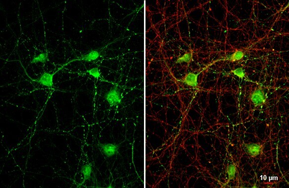
- VGAT Polyclonal Antibody detects VGAT protein by immunofluorescent analysis. Sample: DIV9 rat cortical neuron and Glia cell cells were fixed in 4% paraformaldehyde at RT for 15 min. Green: VGAT stained by VGAT Polyclonal Antibody (Product # PA5-27569) diluted at 1:250. Red: Tau, a Axon marker, stained by Phospho-Tau (Ser262) Polyclonal Antibody [GT287]diluted at 1:500.

- Submitted by
- Invitrogen Antibodies (provider)
- Main image

- Experimental details
- Immunocytochemistry-Immunofluorescence analysis of VGAT was performed in Cultured rat E18 primary cortical neuron, DIV 8. Cells fixed in 4% paraformaldehyde at RT for 15 min. Green: VGAT Polyclonal Antibody (Product # PA5-27569) diluted at 1:250. Red: beta Tubulin 3/ TUJ1, stained by beta Tubulin 3/ TUJ1 antibody. Blue: Fluoroshield with DAPI.

Supportive validation
- Submitted by
- Invitrogen Antibodies (provider)
- Main image

- Experimental details
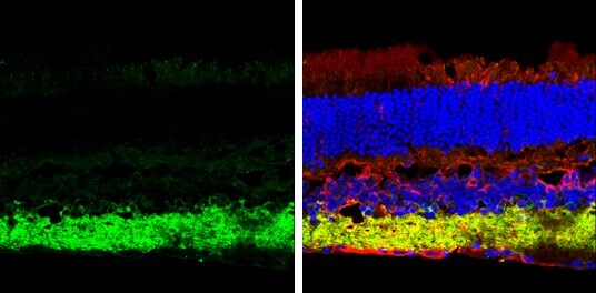
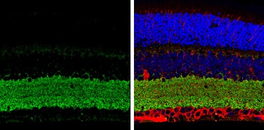
- Immunohistochemistry (Frozen) analysis of VGAT was performed in frozen sectioned adult mouse retina tissue using VGAT Polyclonal Antibody (Product # PA5-27569) at a dilution of 1:250 (Green). Red: beta Tubulin 3/ TUJ1, stained by beta Tubulin 3/ TUJ1 antibody diluted at 1:250. Blue: Fluoroshield with DAPI.
- Submitted by
- Invitrogen Antibodies (provider)
- Main image
- Experimental details
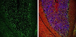
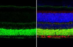
- Immunohistochemistry (Frozen) analysis of VGAT was performed in frozen-sectioned adult mouse cerebellum tissue using VGAT Polyclonal Antibody (Product # PA5-27569) at a dilution of 1:250 (Green). Red: beta Tubulin 3/ TUJ1, stained by beta Tubulin 3/ TUJ1 antibody diluted at 1:500. Blue: Fluoroshield with DAPI.
- Submitted by
- Invitrogen Antibodies (provider)
- Main image
- Experimental details
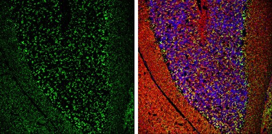
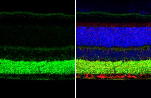
- Immunohistochemistry (Paraffin) analysis of VGAT was performed in paraffin-Embedded adult mouse retina tissue using Green: CRABP2 Polyclonal Antibody (Product # PA5-27451) at a dilution of 1:250. Red: beta Tubulin 3/ TUJ1, stained by beta Tubulin 3/ TUJ1 antibody diluted at 1:500. Blue: Fluoroshield with DAPI.
- Submitted by
- Invitrogen Antibodies (provider)
- Main image
- Experimental details
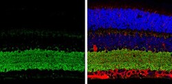
- VGAT Polyclonal Antibody detects VGAT protein at cytoplasm by immunohistochemical analysis. Sample: Paraffin-embedded mouse eye. Green: VGAT stained by VGAT Polyclonal Antibody (Product # PA5-27569) diluted at 1:250. Red: beta Tubulin 3/ Tuj1, a cytoskeleton marker, stained by beta Tubulin 3/ Tuj1 antibody [GT11710] diluted at 1:500. Blue: Fluoroshield with DAPI. Antigen Retrieval: Citrate buffer, pH 6.0, 15 min.
- Submitted by
- Invitrogen Antibodies (provider)
- Main image
- Experimental details
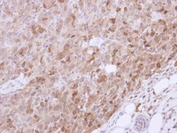
- Immunohistochemical analysis of paraffin-embedded CL1-5 xenograft, using VGAT (Product # PA5-27569) antibody at 1:500 dilution. Antigen Retrieval: EDTA based buffer, pH 8.0, 15 min.

Supportive validation
- Submitted by
- Invitrogen Antibodies (provider)
- Main image
- Experimental details
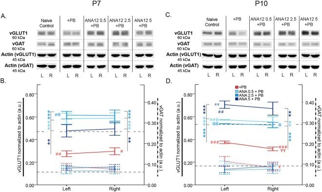
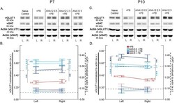
- Figure 7 vGLUT1 vs. vGAT expression 24 h post-ischemia. ( A ) Representative Western blots showing vGLUT1 and vGAT expression at P7. The blots presented for each group were cropped from separate gels for improvement in clarity and conciseness of presentation. Vertical solid black lines delineate the separate blots between groups, while horizontal dotted black lines delineate separate proteins run on different gels for the same sample. All gels were run in the same experimental conditions (see methods for details). (Full-length blots of each tested presented in Supplementary Figure 7 ). ( B ) Quantification of Western blots shown in A. vGLUT1 is downregulated by ischemia and rescued by all doses of ANA12 + PB in a dose-dependent manner. vGAT is not modulated by ischemia. ( C ) Representative Western blots showing vGLUT1 and vGAT expression at P10. ( D ) Quantification of blots shown in C. vGLUT1 is downregulated by ischemia and rescued in a dose-dependent manner by ANA12 + PB. vGAT is unaffected by ischemia. Significance to PB alone: * P < 0.05, ** P < 0.01, *** P < 0.001. Significance to naive control: # P < 0.05, ## P < 0.01, ### P < 0.001. Significance to 5 mg/kg of ANA12 + PB: @ P < 0.05, @@ P < 0.01, @@@ P < 0.001. Ipsilateral to contralateral: gamma P < 0.05, gammagamma P < 0.01, gammagammagamma P < 0.001.