Explore
 Validate
 Learn
Learn Immunohistochemistry
Antibody data
- Antibody Data
- Antigen structure
- References [5]
- Comments [0]
- Validations
- Immunohistochemistry [1]
- Flow cytometry [1]
- Other assay [2]
Submit
Validation data
Reference
Comment
Report error
- Product number
- MA5-11642 - Provider product page

- Provider
- Invitrogen Antibodies
- Product name
- CD42b Monoclonal Antibody (42C01)
- Antibody type
- Monoclonal
- Antigen
- Recombinant full-length protein
- Description
- MA5-11642 targets CD42b in IHC (P) and FACS applications and shows reactivity with Human samples.
- Antibody clone number
- 42C01
- Concentration
- 0.2 mg/mL
Submitted references Clot composition characterization using diffuse reflectance spectroscopy in acute ischemic stroke.
High Mobility Group Box 1 Protein in Cerebral Thromboemboli.
Structural analysis of ischemic stroke thrombi: histological indications for therapy resistance.
Redox control of β2-glycoprotein I-von Willebrand factor interaction by thioredoxin-1.
Crystal structure of a platelet-agglutinating factor isolated from the venom of Taiwan habu (Trimeresurus mucrosquamatus).
Skyrman S, Burström G, Aspegren O, Babic D, Lucassen G, Edström E, Arnberg F, Ohlsson M, Mueller M, Elmi-Terander A, Andersson T
Biomedical optics express 2022 Jun 1;13(6):3311-3323
Biomedical optics express 2022 Jun 1;13(6):3311-3323
High Mobility Group Box 1 Protein in Cerebral Thromboemboli.
Essig F, Babilon L, Vollmuth C, Kollikowski AM, Pham M, Solymosi L, Haeusler KG, Kraft P, Stoll G, Schuhmann MK
International journal of molecular sciences 2021 Oct 19;22(20)
International journal of molecular sciences 2021 Oct 19;22(20)
Structural analysis of ischemic stroke thrombi: histological indications for therapy resistance.
Staessens S, Denorme F, Francois O, Desender L, Dewaele T, Vanacker P, Deckmyn H, Vanhoorelbeke K, Andersson T, De Meyer SF
Haematologica 2020;105(2):498-507
Haematologica 2020;105(2):498-507
Redox control of β2-glycoprotein I-von Willebrand factor interaction by thioredoxin-1.
Passam FH, Rahgozar S, Qi M, Raftery MJ, Wong JW, Tanaka K, Ioannou Y, Zhang JY, Gemmell R, Qi JC, Giannakopoulos B, Hughes WE, Hogg PJ, Krilis SA
Journal of thrombosis and haemostasis : JTH 2010 Aug;8(8):1754-62
Journal of thrombosis and haemostasis : JTH 2010 Aug;8(8):1754-62
Crystal structure of a platelet-agglutinating factor isolated from the venom of Taiwan habu (Trimeresurus mucrosquamatus).
Huang KF, Ko TP, Hung CC, Chu J, Wang AH, Chiou SH
The Biochemical journal 2004 Mar 1;378(Pt 2):399-407
The Biochemical journal 2004 Mar 1;378(Pt 2):399-407
No comments: Submit comment
Supportive validation
- Submitted by
- Invitrogen Antibodies (provider)
- Main image
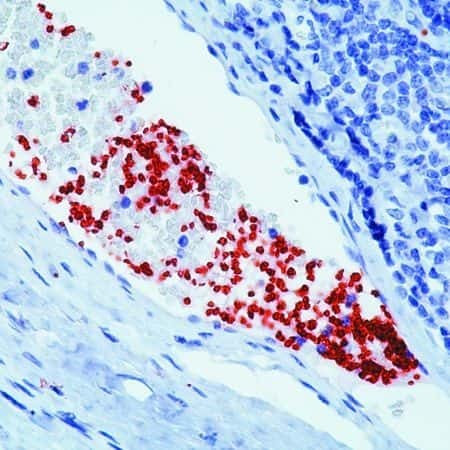
- Experimental details
- Formalin-fixed, paraffin-embedded human tonsil stained with CD42b antibody using peroxidase-conjugate and AEC chromogen. Note staining of platelets in the blood vessels.
Supportive validation
- Submitted by
- Invitrogen Antibodies (provider)
- Main image
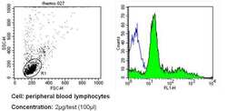
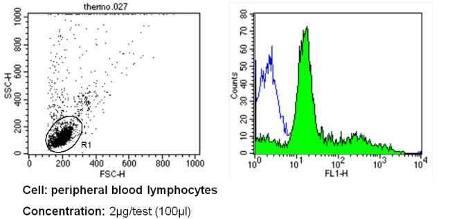
- Experimental details
- Flow cytometry analysis of CD42b in PBMC cells (green) compared to an isotype control (blue). Human blood was collected, combined with a hydrophilic polysaccharide, centrifuged, transferred to a conical tube and washed with PBS. 50 µL of cell solution was added to each tube at a dilution of 2x10^7 cells/mL, followed by the addition of 50 µL of isotype control and primary antibody (Product # MA5-11642) at a dilution of 2 µg/test. Cells were incubated for 30 min at 4ºC and washed with a cell buffer, followed by incubation with a DyLight 488-conjugated secondary antibody for 30 min at 4ºC in the dark. FACS analysis was performed using 400 µL of cell buffer.
Supportive validation
- Submitted by
- Invitrogen Antibodies (provider)
- Main image
- Experimental details
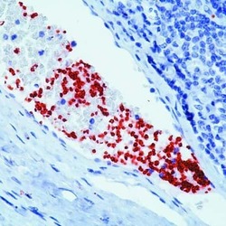
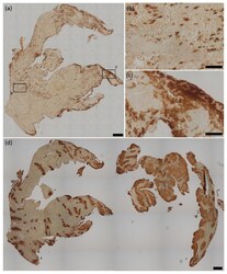
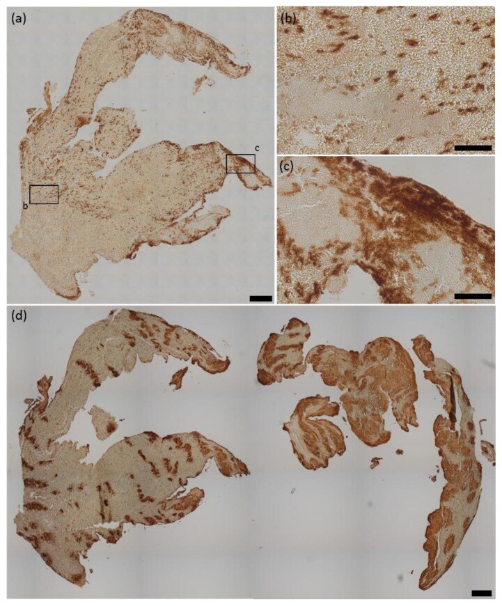
- Figure 1 HMGB1 and platelets in cerebral thromboemboli. ( a ) Representative HMGB1 stained thromboemboli and magnifications illustrating the presence of ( b ) small, spotted HMGB1 regions, whereas, especially in peripheral regions, ( c ) extensive areas of HMGB1 were found. ( d ) Representative CD42b stained thromboemboli illustrating the diversity of platelet-rich areas within cerebral thromboemboli. HMGB1 = high mobility group box 1. Scale bars: ( a , d ) 200 um, ( b , c ) 50 um.
- Submitted by
- Invitrogen Antibodies (provider)
- Main image

- Experimental details
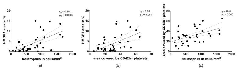
- Figure 4 Correlation between ( a ) the area of HMGB1 (in % of the total thromboembolus area) and the amount of neutrophils, ( b ) the area of HMGB1 and the area covered by CD42b-positive platelets, and ( c ) the amount of neutrophils with the area covered by CD42b-positive platelets. p = level of significance. r = correlation coefficient. s = Spearman, p = Pearson.