 Explore
 Validate
 Learn
Learn Immunocytochemistry
Antibody data
- Antibody Data
- Antigen structure
- References [0]
- Comments [0]
- Validations
- Immunocytochemistry [3]
Submit
Validation data
Reference
Comment
Report error
- Product number
- MA1-16573 - Provider product page

- Provider
- Invitrogen Antibodies
- Product name
- Separase Monoclonal Antibody (XJ11-4D7)
- Antibody type
- Monoclonal
- Antigen
- Other
- Reactivity
- Human, Mouse
- Host
- Mouse
- Isotype
- IgG
- Antibody clone number
- XJ11-4D7
- Vial size
- 100 μL
- Concentration
- Conc. Not Determined
- Storage
- -20°C, Avoid Freeze/Thaw Cycles
No comments: Submit comment
Supportive validation
- Submitted by
- Invitrogen Antibodies (provider)
- Main image

- Experimental details
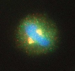
- Immunocytochemistry analysis of Separase in HeLa cells. Samples were incubated in Separase monoclonal antibody (Product # MA1-16573). Overlay [blue] of centrosomal staining. Centrosomal staining of separase [yellow] and gamma tubulin [green] in mitotic-metaphase cells and nuclear staining of separase in pre-mitotic cells.

- Submitted by
- Invitrogen Antibodies (provider)
- Main image

- Experimental details
- Immunocytochemistry analysis of Separase in HeLa cells. Samples were incubated in Separase monoclonal antibody (Product # MA1-16573). Overlay [blue] of centrosomal staining. Centrosomal staining of separase [yellow] and gamma tubulin [green] in mitotic-metaphase cells and nuclear staining of separase in pre-mitotic cells.

- Submitted by
- Invitrogen Antibodies (provider)
- Main image
- Experimental details
- Immunocytochemistry analysis of Separase in HeLa cells. Samples were incubated in Separase monoclonal antibody (Product # MA1-16573). Overlay [blue] of centrosomal staining. Centrosomal staining of separase [yellow] and gamma tubulin [green] in mitotic-metaphase cells and nuclear staining of separase in pre-mitotic cells.
