 Explore
 Validate
 Learn
Learn Western blot
 Immunocytochemistry
Antibody data
- Antibody Data
- Antigen structure
- References [0]
- Comments [0]
- Validations
- Western blot [1]
- Immunohistochemistry [1]
- Other assay [1]
Submit
Validation data
Reference
Comment
Report error
- Product number
- NBP2-25166 - Provider product page

- Provider
- Novus Biologicals
- Product name
- Mouse Monoclonal visinin-like 1 Antibody
- Antibody type
- Monoclonal
- Description
- Immunogen affinity purified.
- Reactivity
- Human, Mouse, Rat
- Host
- Mouse
- Isotype
- IgG
- Vial size
- 0.1 ml
- Concentration
- 1 mg/ml
- Storage
- Store at 4C short term. Aliquot and store at -20C long term. Avoid freeze-thaw cycles.
No comments: Submit comment
Supportive validation
- Submitted by
- Novus Biologicals (provider)
- Main image
- Experimental details
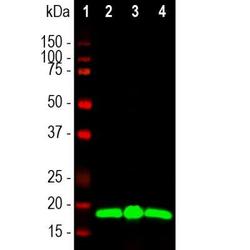
- Western Blot: visinin-like 1 Antibody (2D11) [NBP2-25166] - Western blot analysis of different tissue lysates using mouse mAb to Visinin-like Protein 1 (VLP1), NBP2-25166, dilution 1:1,000 in green: [1] protein standard (red), [2] rat brain [3] rat cerebellum, and [4] mouse brain. The band at around 20kDa mark corresponds to VLP1 protein.

Supportive validation
- Submitted by
- Novus Biologicals (provider)
- Main image
- Experimental details
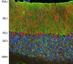
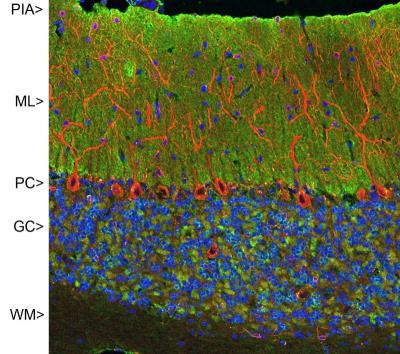
- Immunohistochemistry: visinin-like 1 Antibody (2D11) [NBP2-25166] - Confocal image of adult rat cerebellar cortex stained with NBP2-25166 (green), chicken polyclonal antibody to MAP2 NB300-213 (red) and DNA (blue). NBP2-25166 reveals synapses in the molecular layer (ML) strongly. Synaptic regions are also seen in the granule cell layer (GC). The perikarya of Purkinje cells (PC) are revealed with MAP2 antibody. Little staining is seen in the white matter (WM).
Supportive validation
- Submitted by
- Novus Biologicals (provider)
- Main image
- Experimental details
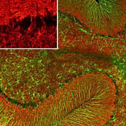
- Immunohistochemistry Free-Floating: visinin-like 1 Antibody (2D11) [NBP2-25166] - Immunofluorescent analysis of rat cerebellum section stained with mouse mAb to VLP1, NBP2-25166, dilution 1:500, in red and costained with rabbit pAb to GFAP, dilution 1:5,000 in green. The blue is DAPI staining of nuclear DNA. Following transcardial perfusion of rat with 4% paraformaldehyde, brain was post fixed for 24 hours, cut to 45uM, and free-floating sections were stained with the above antibodies. The VLP1 antibody reveals protein expressed in granule cell membranes and in synapses in the white matter and molecular layers of the cerebellum. The GFAP antibody stains the processes of Bergmann glia and astroglia.
