Explore
 Validate
 Learn
Learn Western blot
 Immunocytochemistry
Antibody data
- Antibody Data
- Antigen structure
- References [0]
- Comments [0]
- Validations
- Western blot [2]
- Other assay [1]
Submit
Validation data
Reference
Comment
Report error
- Product number
- NBP2-25169 - Provider product page

- Provider
- Novus Biologicals
- Product name
- Rabbit Polyclonal visinin-like 1 Antibody
- Antibody type
- Polyclonal
- Description
- Immunogen affinity purified.
- Reactivity
- Human, Mouse, Rat
- Host
- Rabbit
- Isotype
- IgG
- Vial size
- 0.1 ml
- Concentration
- 1 mg/ml
- Storage
- Store at 4C short term. Aliquot and store at -20C long term. Avoid freeze-thaw cycles.
No comments: Submit comment
Supportive validation
- Submitted by
- Novus Biologicals (provider)
- Main image
- Experimental details
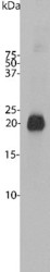
- Western Blot: visinin-like 1 Antibody [NBP2-25169] - Bovine cerebellum homogenate. Note the strong clean band running at 22 kDa.

- Submitted by
- Novus Biologicals (provider)
- Main image

- Experimental details
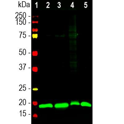
- Western Blot: visinin-like 1 Antibody [NBP2-25169] - Analysis of different tissue lysates using rabbit pAb to visinin-like Protein 1 (VLP1), NBP2-25169, dilution 1:20,000 in green: [1] protein standard (red), [2] rat brain [3] mouse brain, [4] pig hippocampus, and [5] cow cerebellum. The band at ~20kDa corresponds to the VLP1 protein.
Supportive validation
- Submitted by
- Novus Biologicals (provider)
- Main image
- Experimental details
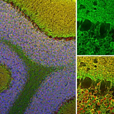
- Immunohistochemistry Free-Floating: visinin-like 1 Antibody [NBP2-25169] - Analysis of rat cerebellum section stained with rabbit pAb to VLP1, NBP2-25169, dilution 1:2,000 in green, and costained with mouse mAb to calretinin, dilution 1:2,000 in red. The blue is DAPI staining of nuclear DNA. Following transcardial perfusion of rat with 4% paraformaldehyde, brain was post fixed for 24 hours, cut to 45uM, and free-floating sections were stained with the above antibodies. The VLP1 antibody reveals protein expressed in granule cells membranes and their synapses in both the granular and molecular layer of the cerebellum. The calretinin antibody stains the cytoplasm of neurons in the nuclear and molecular layers of cerebellum.