Explore
 Validate
 Learn
Learn Western blot
 Immunocytochemistry
Antibody data
- Antibody Data
- Antigen structure
- References [0]
- Comments [0]
- Validations
- Western blot [2]
- Immunohistochemistry [2]
Submit
Validation data
Reference
Comment
Report error
- Product number
- LS-B10380 - Provider product page

- Provider
- LSBio
- Product name
- IHC-plus™ VILIP / VSNL1 Antibody (clone 2D11) LS-B10380
- Antibody type
- Monoclonal
- Description
- Affinity purified
- Reactivity
- Human, Mouse, Rat, Bovine
- Host
- Mouse
- Isotype
- IgG
- Antibody clone number
- 2D11
- Storage
- Store at 4°C or -20°C. Avoid freeze-thaw cycles.
No comments: Submit comment
Enhanced validation
- Submitted by
- LSBio (provider)
- Enhanced method
- Genetic validation
- Main image
- Experimental details
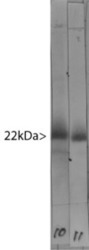
- Western blot of bovine cerebellum homogenate stained with VILIP / VSNL1 antibody in lane 10. Note the strong clean band running at 22kDa. Lane 11 shows the same material stained with an alternate antibody to VSNL1, which binds to the same band.

- Submitted by
- LSBio (provider)
- Enhanced method
- Genetic validation
- Main image
- Experimental details
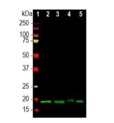
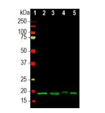
- Western blot using antibody at a dilution of 1:1000. Lane 1, marker; Lane 2, rat brain; Lane 3, mouse brain; Lane 4, pig hippocampus; and Lane 5, bovine cerebellum.
Enhanced validation
- Submitted by
- LSBio (provider)
- Enhanced method
- Genetic validation
- Main image
- Experimental details
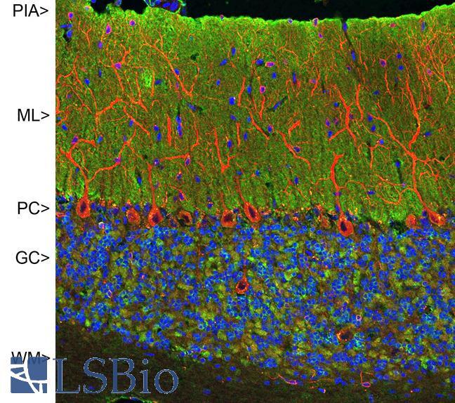
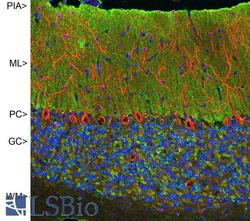
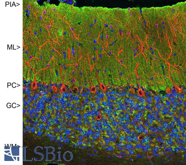
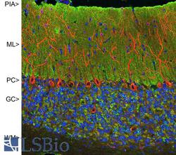
- Confocal image of adult rat cerebellar cortex stained with VILIP / VSNL1 antibody (green), chicken polyclonal antibody to MAP2 (red) and DNA (blue). The VILIP / VSNL1 antibody antibody reveals synapses in the molecular layer (ML) strongly. Synaptic regions are also seen in the granule cell layer (GC). The perikarya of Purkinje cells (PC) are revealed with MAP2 antibody (4). Little staining is seen in the white matter (WM).
- Submitted by
- LSBio (provider)
- Enhanced method
- Genetic validation
- Main image
- Experimental details
- Confocal image of adult rat cerebellar cortex stained with VILIP / VSNL1 antibody (green), chicken polyclonal antibody to MAP2 (red) and DNA (blue). The VILIP / VSNL1 antibody antibody reveals synapses in the molecular layer (ML) strongly. Synaptic regions are also seen in the granule cell layer (GC). The perikarya of Purkinje cells (PC) are revealed with MAP2 antibody (4). Little staining is seen in the white matter (WM).