 Explore
 Validate
 Learn
Learn Western blot
 Immunocytochemistry
 Immunohistochemistry
Antibody data
- Antibody Data
- Antigen structure
- References [0]
- Comments [0]
- Validations
- Western blot [1]
- Immunocytochemistry [3]
Submit
Validation data
Reference
Comment
Report error
- Product number
- LS-B10407 - Provider product page

- Provider
- LSBio
- Product name
- IHC-plus™ LAMP1 / CD107a Antibody (clone 6E2) LS-B10407
- Antibody type
- Monoclonal
- Description
- Affinity purified
- Reactivity
- Human, Mouse, Rat, Bovine, Porcine
- Host
- Mouse
- Isotype
- IgG
- Antibody clone number
- 6E2
- Storage
- Store at 4°C or -20°C. Avoid freeze-thaw cycles.
No comments: Submit comment
Enhanced validation
- Submitted by
- LSBio (provider)
- Enhanced method
- Genetic validation
- Main image
- Experimental details
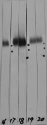
- Western strip blots of HeLa cell crude extracts with two different preparations of CD107a / LAMP1 antibody in strips 17 and 18. Both preparations bind to a diffuse band running at between ~90kDa and ~120kDa as expected, and show no appreciably cross reactivity with any other protein.

Supportive validation
- Submitted by
- LSBio (provider)
- Enhanced method
- Genetic validation
- Main image
- Experimental details
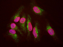
- HeLa cells staining with CD107a / LAMP1 antibody (green), and counterstained with chicken polyclonal antibody to Lamin A/C (red) and DNA (blue). The CD107a / LAMP1 antibody antibody reveals strong cytoplasmic staining of lysosomes and early endosomes, while the Lamin A/C antibody binds to the nuclear lamina. Since both DNA (blue) and Lamin A/C (red) are associated with the nuclear compartment, this region appears crimson in this image.

- Submitted by
- LSBio (provider)
- Main image

- Experimental details
- HeLa cells staining with CD107a / LAMP1 antibody (green), and counterstained with chicken polyclonal antibody to Lamin A/C (red) and DNA (blue). The CD107a / LAMP1 antibody antibody reveals strong cytoplasmic staining of lysosomes and early endosomes, while the Lamin A/C antibody binds to the nuclear lamina. Since both DNA (blue) and Lamin A/C (red) are associated with the nuclear compartment, this region appears crimson in this image.
