 Explore
 Validate
 Learn
Learn Western blot
 Immunohistochemistry
Antibody data
- Antibody Data
- Antigen structure
- References [2]
- Comments [0]
- Validations
- Western blot [1]
- Immunocytochemistry [1]
Submit
Validation data
Reference
Comment
Report error
- Product number
- ABIN1580431 - Provider product page

- Provider
- antibodies-online
- Product name
- anti-Lysosomal-Associated Membrane Protein 1 (LAMP1) antibody
- Antibody type
- Monoclonal
- Antigen
- Other
- Description
- affinity purified antibody
- Reactivity
- Human, Mouse, Rat, Bovine, Porcine
- Host
- Mouse
- Isotype
- IgG
- Antibody clone number
- 5H6
- Vial size
- 100 μL
- Concentration
- 1 mg/mL
- Storage
- Store at 4°C short term or -20°C long term.
- Handling
- Avoid repeated freezing and thawing.
Submitted references Derived protein sequence, oligosaccharides, and membrane insertion of the 120-kDa lysosomal membrane glycoprotein (lgp120): identification of a highly conserved family of lysosomal membrane glycoproteins.
Translocation and clustering of endosomes and lysosomes depends on microtubules.
Howe CL, Granger BL, Hull M, Green SA, Gabel CA, Helenius A, Mellman I
Proceedings of the National Academy of Sciences of the United States of America 1988 Oct;85(20):7577-81
Proceedings of the National Academy of Sciences of the United States of America 1988 Oct;85(20):7577-81
Translocation and clustering of endosomes and lysosomes depends on microtubules.
Matteoni R, Kreis TE
The Journal of cell biology 1987 Sep;105(3):1253-65
The Journal of cell biology 1987 Sep;105(3):1253-65
No comments: Submit comment
Supportive validation
- Submitted by
- antibodies-online (provider)
- Main image
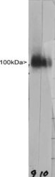
- Experimental details
- Western strip blots of HeLa cell crude extracts stained with anti LAMP1 antibody ABIN1580431 in strip 9. Lane 10 shows staining with our other LAMP1 antibody MCA-6E2. Both antibodies bind to a diffuse band running at between ~90 and ~120 kDa as expected, and show no appreciably cross reactivity with any other protein.

Supportive validation
- Submitted by
- antibodies-online (provider)
- Main image
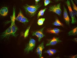
- Experimental details
- HeLa cells staining with ABIN1580431 (red), and counterstained with chicken polyclonal antibody to Vimentin CPCA-Vim (green) and DNA (blue). The ABIN1580431 antibody reveals strong punctate cytoplasmic staining corresponding to lysosomes and late endosomes, while the Vimentin antibody reveals cytoplasmic intermediate filaments.
