Explore
 Validate
 Learn
Learn Western blot
 Flow cytometry
Antibody data
- Antibody Data
- Antigen structure
- References [3]
- Comments [0]
- Validations
- Western blot [1]
- Immunocytochemistry [1]
- Immunohistochemistry [1]
Submit
Validation data
Reference
Comment
Report error
- Product number
- MAB4800 - Provider product page

- Provider
- Novus Biologicals
- Product name
- Mouse Monoclonal LAMP-1/CD107a Antibody
- Antibody type
- Monoclonal
- Description
- Protein A or G purified from hybridoma culture supernatant. Detects human LAMP1/CD107a in direct ELISAs.
- Reactivity
- Human
- Host
- Mouse
- Conjugate
- Unconjugated
- Isotype
- IgG
- Vial size
- 100 ug
- Concentration
- LYOPH
- Storage
- Use a manual defrost freezer and avoid repeated freeze-thaw cycles. 12 months from date of receipt, -20 to -70 degreesC as supplied. 1 month, 2 to 8 degreesC under sterile conditions after reconstitution. 6 months, -20 to -70 degreesC under sterile conditions after reconstitution.
Submitted references Sorting Nexin 9 facilitates podocin endocytosis in the injured podocyte.
Role of OSGIN1 in mediating smoking-induced autophagy in the human airway epithelium.
APOL1 variants change C-terminal conformational dynamics and binding to SNARE protein VAMP8.
Sasaki Y, Hidaka T, Ueno T, Akiba-Takagi M, Oliva Trejo JA, Seki T, Nagai-Hosoe Y, Tanaka E, Horikoshi S, Tomino Y, Suzuki Y, Asanuma K
Scientific reports 2017 Mar 7;7:43921
Scientific reports 2017 Mar 7;7:43921
Role of OSGIN1 in mediating smoking-induced autophagy in the human airway epithelium.
Wang G, Zhou H, Strulovici-Barel Y, Al-Hijji M, Ou X, Salit J, Walters MS, Staudt MR, Kaner RJ, Crystal RG
Autophagy 2017 Jul 3;13(7):1205-1220
Autophagy 2017 Jul 3;13(7):1205-1220
APOL1 variants change C-terminal conformational dynamics and binding to SNARE protein VAMP8.
Madhavan SM, O'Toole JF, Konieczkowski M, Barisoni L, Thomas DB, Ganesan S, Bruggeman LA, Buck M, Sedor JR
JCI insight 2017 Jul 20;2(14)
JCI insight 2017 Jul 20;2(14)
No comments: Submit comment
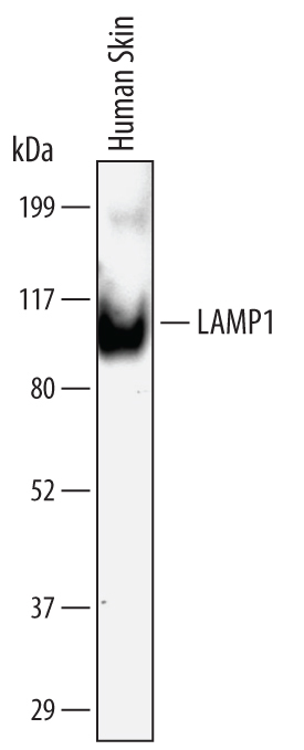
Supportive validation
- Submitted by
- Novus Biologicals (provider)
- Main image

- Experimental details
- Detection of Human LAMP1/CD107a by Western Blot. Western blot shows lysates of human skin tissue. PVDF Membrane was probed with 2 µg/mL of Mouse Anti-Human LAMP1/CD107a Monoclonal Antibody (Catalog # MAB4800) followed by HRP-conjugated Anti-Mouse IgG Secondary Antibody (Catalog # HAF007). A specific band was detected for LAMP1/CD107a at approximately 100 kDa (as indicated). This experiment was conducted under non-reducing conditions and using Immunoblot Buffer Group 1.
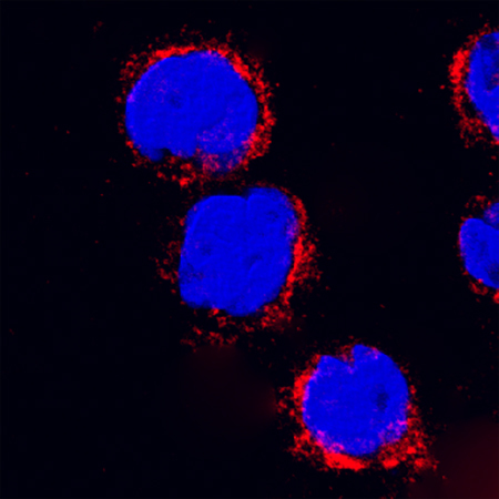
Supportive validation
- Submitted by
- Novus Biologicals (provider)
- Main image

- Experimental details
- LAMP-1/CD107a in THP-1 Human Cell Line. LAMP-1/CD107a was detected in immersion fixed THP-1 human acute monocytic leukemia cell line using Mouse Anti-Human LAMP-1/CD107a Monoclonal Antibody (Catalog # MAB4800) at 25 µg/mL for 3 hours at room temperature. Cells were stained using the NorthernLights™ 557-conjugated Anti-Mouse IgG Secondary Antibody (red; Catalog # NL007) and counterstained with DAPI(blue). Specific staining was localized to cytoplasmic. View our protocol for Fluorescent ICC Staining of Cells on Coverslips.
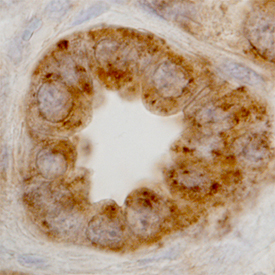
Supportive validation
- Submitted by
- Novus Biologicals (provider)
- Main image

- Experimental details
- LAMP1/CD107a in Human Kidney. LAMP1/CD107a was detected in immersion fixed paraffin-embedded sections of human kidney using Mouse Anti-Human LAMP1/CD107a Monoclonal Antibody (Catalog # MAB4800) at 15 µg/mL overnight at 4 °C. Before incubation with the primary antibody, tissue was subjected to heat-induced epitope retrieval using Antigen Retrieval Reagent-Basic (Catalog # CTS013). Tissue was stained using the Anti-Mouse HRP-DAB Cell & Tissue Staining Kit (brown; Catalog # CTS002) and counterstained with hematoxylin (blue). Specific staining was localized to lysosomes in epithelial cells. View our protocol for Chromogenic IHC Staining of Paraffin-embedded Tissue Sections.