 Explore
 Validate
 Learn
Learn Western blot
 Immunocytochemistry
Antibody data
- Antibody Data
- Antigen structure
- References [2]
- Comments [0]
- Validations
- Immunocytochemistry [2]
- Flow cytometry [1]
Submit
Validation data
Reference
Comment
Report error
- Product number
- MA5-18121 - Provider product page

- Provider
- Invitrogen Antibodies
- Product name
- LAMP1 Monoclonal Antibody (H4A3), Alexa Fluor™ 488
- Antibody type
- Monoclonal
- Antigen
- Other
- Description
- This antibody recognizes an extracellular/luminal epitope of CD107a, an approximately 100-120 kDa glycoprotein expressed mainly on lysosomal, but also on the plasma membrane.
- Reactivity
- Human, Mouse
- Host
- Mouse
- Conjugate
- Green dye
- Isotype
- IgG
- Antibody clone number
- H4A3
- Vial size
- 100 Tests
- Storage
- 4° C, store in dark, DO NOT FREEZE!
Submitted references Elevated glucosylsphingosine in Gaucher disease induced pluripotent stem cell neurons deregulates lysosomal compartment through mammalian target of rapamycin complex 1.
Quantification of natural killer cell polarization and visualization of synaptic granule externalization by imaging flow cytometry.
Srikanth MP, Jones JW, Kane M, Awad O, Park TS, Zambidis ET, Feldman RA
Stem cells translational medicine 2021 Jul;10(7):1081-1094
Stem cells translational medicine 2021 Jul;10(7):1081-1094
Quantification of natural killer cell polarization and visualization of synaptic granule externalization by imaging flow cytometry.
Viswanath DI, Mace EM, Hsu HT, Orange JS
Clinical immunology (Orlando, Fla.) 2017 Apr;177:70-75
Clinical immunology (Orlando, Fla.) 2017 Apr;177:70-75
No comments: Submit comment
Supportive validation
- Submitted by
- Invitrogen Antibodies (provider)
- Main image

- Experimental details
- Immunofluorescence analysis of LAMP1 was performed using 70% confluent log phase HeLa cells. The cells were fixed with 4% paraformaldehyde for 10 minutes, permeabilized with 0.1% Triton™ X-100 for 10 minutes, and blocked with 1% BSA for 1 hour at room temperature. The cells were labeled with LAMP1, Alexa Fluor 488, Mouse Monoclonal antibody (Product # MA5-18121) at 1:100 dilution in 0.1% BSA and incubated at 4 degree Celsius overnight (Panel a: green). Nuclei (Panel b: blue) were stained with SlowFade® Gold Antifade Mountant with DAPI (Product # S36938). F-actin (Panel c: red) was stained with Rhodamine Phalloidin (Product # R415, 1:300). Panel d represents the merged image showing cytoplasmic localization. Panel e represents the isotype control. The images were captured at 60X magnification.
- Conjugate
- Green dye

- Submitted by
- Invitrogen Antibodies (provider)
- Main image
- Experimental details
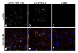
- Knockdown of LAMP1 was achieved by transfecting HeLa cells with LAMP1 specific siRNAs (Silencer® select Product # s8080, s8081 ). Immunofluorescence analysis was performed using untransfected HeLa cells (panels a, d), transfected with non-specific scrambled siRNA (panels b,e) and transfected with LAMP1 specific siRNAs (panel c,f). Cells were fixed, permeabilized, and probed with LAMP1 Monoclonal Antibody (H4A3), Alexa Fluor 488 (Product # MA5-18121, 1:250 dilution). Nuclei (blue) were stained using ProLong™ Diamond Antifade Mountant with DAPI (Product # P36962) and Rhodamine Phalloidin (Product # R415, 1:300) was used for cytoskeletal F-actin (red) staining. Reduction of specific cytoplasmic localization was observed upon siRNA mediated knockdown (panel c,f) confirming specificity of the antibody to LAMP1. The images were captured at 60X magnification.
- Conjugate
- Green dye

Supportive validation
- Submitted by
- Invitrogen Antibodies (provider)
- Main image

- Experimental details
- Flow cytometry analysis (intracellular staining) of human peripheral blood cells with anti-CD107a (H4A3) Alexa Fluor® 488 Monoclonal antibody (Product # MA5-18121).
- Conjugate
- Green dye
