 Explore
 Validate
 Learn
Learn Western blot
Antibody data
- Antibody Data
- Antigen structure
- References [2]
- Comments [0]
- Validations
- Western blot [1]
- Immunocytochemistry [1]
- Flow cytometry [1]
Submit
Validation data
Reference
Comment
Report error
- Product number
- AF4320 - Provider product page

- Provider
- R&D Systems
- Product name
- Mouse LAMP-1/CD107a Lumenal Domain Antibody
- Antibody type
- Polyclonal
- Description
- Antigen Affinity-purified. Detects mouse LAMP1/CD107a Lumenal Domain in direct ELISAs and Western blots. In direct ELISAs, less than 10% cross-reactivity with recombinant human LAMP1 is observed.
- Reactivity
- Mouse
- Host
- Goat
- Conjugate
- Unconjugated
- Antigen sequence
P11438- Isotype
- IgG
- Vial size
- 100 ug
- Concentration
- LYOPH
- Storage
- Use a manual defrost freezer and avoid repeated freeze-thaw cycles. 12 months from date of receipt, -20 to -70 °C as supplied. 1 month, 2 to 8 °C under sterile conditions after reconstitution. 6 months, -20 to -70 °C under sterile conditions after reconstitution.
Submitted references Cystinosin, the small GTPase Rab11, and the Rab7 effector RILP regulate intracellular trafficking of the chaperone-mediated autophagy receptor LAMP2A.
Purkinje Cells Are More Vulnerable to the Specific Depletion of Cathepsin D Than to That of Atg7.
Zhang J, Johnson JL, He J, Napolitano G, Ramadass M, Rocca C, Kiosses WB, Bucci C, Xin Q, Gavathiotis E, Cuervo AM, Cherqui S, Catz SD
The Journal of biological chemistry 2017 Jun 23;292(25):10328-10346
The Journal of biological chemistry 2017 Jun 23;292(25):10328-10346
Purkinje Cells Are More Vulnerable to the Specific Depletion of Cathepsin D Than to That of Atg7.
Koike M, Shibata M, Sunabori T, Yamaguchi J, Sakimura K, Komatsu M, Tanaka K, Uchiyama Y
The American journal of pathology 2017 Jul;187(7):1586-1600
The American journal of pathology 2017 Jul;187(7):1586-1600
No comments: Submit comment
Supportive validation
- Submitted by
- R&D Systems (provider)
- Main image
- Experimental details
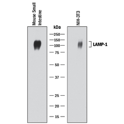
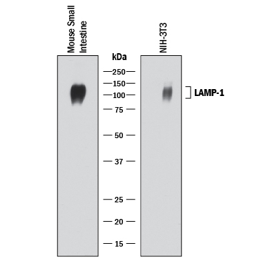
- Detection of Mouse LAMP-1/CD107a by Western Blot. Western blot shows lysates of mouse small intestine tissue and NIH-3T3 mouse embryonic fibroblast cell line. PVDF membrane was probed with 0.2 µg/mL of Goat Anti-Mouse LAMP-1/CD107a Lumenal Domain Antigen Affinity-purified Polyclonal Antibody (Catalog # AF4320) followed by HRP-conjugated Anti-Goat IgG Secondary Antibody (Catalog # HAF017). A specific band was detected for LAMP-1/CD107a at approximately 100-120 kDa (as indicated). This experiment was conducted under reducing conditions and using Immunoblot Buffer Group 1.
Supportive validation
- Submitted by
- R&D Systems (provider)
- Main image
- Experimental details
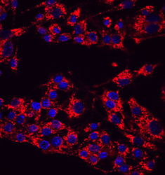
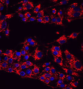
- LAMP1/CD107a in RAW 264.7 Mouse Cell Line. LAMP1/CD107a was detected in immersion fixed RAW 264.7 mouse monocyte/macrophage cell line using Goat Anti-Mouse LAMP1/CD107a Lumenal Domain Antigen Affinity-purified Polyclonal Antibody (Catalog # AF4320) at 10 µg/mL for 3 hours at room temperature. Cells were stained using the NorthernLights™ 557-conjugated Anti-Goat IgG Secondary Antibody (red; Catalog # NL001) and counterstained with DAPI (blue). Specific staining was localized to cytoplasm. View our protocol for Fluorescent ICC Staining of Cells on Coverslips.
Supportive validation
- Submitted by
- R&D Systems (provider)
- Main image
- Experimental details
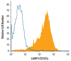
- Detection of LAMP-1 in RAW 264.7 Mouse Cell Line by Flow Cytometry. RAW 264.7 mouse monocyte/macrophage cell line was stained with Goat Anti-Mouse LAMP1/CD107a Lumenal Domain Antigen Affinity-purified Polyclonal Antibody (Catalog # AF4320, filled histogram) or control antibody (Catalog # AB-108-C, open histogram), followed by Allophycocyanin-conjugated Anti-Goat IgG Secondary Antibody (Catalog # F0108). To facilitate intracellular staining, cells were fixed with Flow Cytometry Fixation Buffer (Catalog # FC004) and permeabilized with Flow Cytometry Permeabilization/Wash Buffer I (Catalog # FC005). View our protocol for Staining Intracellular Molecules.
