Explore
 Validate
 Learn
Learn Western blot
 Flow cytometry
Antibody data
- Antibody Data
- Antigen structure
- References [0]
- Comments [0]
- Validations
- Flow cytometry [3]
Submit
Validation data
Reference
Comment
Report error
- Product number
- GTX31229 - Provider product page

- Provider
- GeneTex
- Product name
- LAMP1 antibody [4E9/11]
- Antibody type
- Monoclonal
- Reactivity
- Porcine
- Host
- Mouse
No comments: Submit comment
Supportive validation
- Submitted by
- GeneTex (provider)
- Main image
- Experimental details
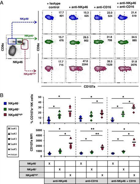
- Published customer image: Mouse anti Pig CD107a antibody, clone 4E9/11 used for the detection of CD107a in porcine NK cells by flow cytometry Image caption: NKp46high NK cells in spleen showed the highest cytolytic capacity. The cytolytic capacity of NKp46-defined NK-cell subsets (CD8a+NKp46-: blue, CD8a+NKp46+: green, CD8adim/-NKp46high: red) isolated from spleen was analysed after receptor-mediated degranulation. Cells were stimulated and gated for flow cytometric analysis as outlined in Figure 4. (A) Numbers indicate the percentage of CD107a+ cells and the mean fluorescence intensity of CD107a within respective NKp46-gates. Results are representative of experiments with five different animals. (B) CD107a expression analyses of five animals analysed. The proportion of CD107a+ cells within the different NKp46-defined subsets is shown in the upper graphs. Percentage of CD107a+ NK cells was calculated by subtracting spontaneous degranulation observed in cultures stimulated with isotype-control antibodies from the frequency of CD107a+ cells in cultures stimulated with NKp46 and/or CD16 mAbs. The lower graphs show the mean fluorescence intensity of CD107a within the respective subsets. Mean values are represented by a black bar. Significant differences between the subsets are indicated (* = p From: Mair KH, Mullebner A, Essler SE, Duvigneau JC, Storset AK, Saalmuller A, Gerner W. Porcine CD8adim/-NKp46high NK cells are in a highly activated state. Vet Res. 2013 Mar 1;44:13 .
- Submitted by
- GeneTex (provider)
- Main image
- Experimental details
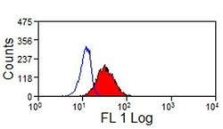
- Pig peripheral blood granulocytes stained with Mouse anti Pig CD107a followed by Goat anti Mouse IgG:FITC . Membrane permeabilisation was achieved with Leucoperm ™

- Submitted by
- GeneTex (provider)
- Main image
- Experimental details
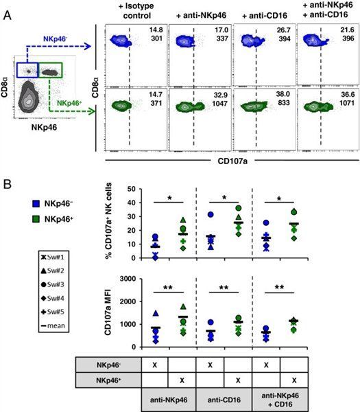
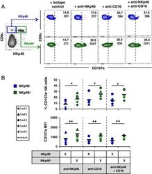
- Published customer image: Mouse anti Pig CD107a antibody, clone 4E9/11 used for the detection of CD107a in porcine NK cells by flow cytometry Image caption: Analysis of degranulation capacity in NKp46-defined NK-cell subsets in blood. The cytolytic capacity of NKp46-defined NK-cell subsets (CD8a+NKp46-: blue, CD8a+NKp46+: green) isolated from blood was analysed after receptor triggering. Cells were stimulated with rhIL-2 and rpIL-15 overnight. Triggering of NK-receptors was performed by using monoclonal antibodies against NKp46, CD16 or a combination of both. Irrelevant isotype-matched antibody served as negative control. NK-cell receptor mediated degranulation was assessed by measuring the expression of CD107a on the cell surface by four-colour flow cytometry after one hour incubation. CD107a expression was measured on CD3- lymphocytes (not shown), followed by gating on the respective NKp46-defined NK subsets. (A) Numbers indicate the percentage of CD107a+ cells and the mean fluorescence intensity of CD107a within respective NKp46-gates. Results are representative of experiments with five different animals. (B) CD107a expression analyses of five animals analysed. The proportion of CD107a+ cells within the different NKp46-defined subsets is shown in the upper graphs. Percentage of CD107a+ NK cells was calculated by subtracting spontaneous degranulation observed in cultures stimulated with isotype-control antibodies from the frequency of CD107a+ cells in cultures stimulated with NKp46 and/or CD16 mAbs. The lower graphs show the mean fluorescence intensity of CD107a within the respective subsets. Mean values are represented by a black bar. Significant differences between the subsets are indicated (* = p From: Mair KH, Mullebner A, Essler SE, Duvigneau JC, Storset AK, Saalmuller A, Gerner W. Porcine CD8adim/-NKp46high NK cells are in a highly activated state. Vet Res. 2013 Mar 1;44:13 .