Explore
 Validate
 Learn
Learn Western blot
Antibody data
- Antibody Data
- Antigen structure
- References [0]
- Comments [0]
- Validations
- Western blot [1]
- Immunocytochemistry [3]
- Immunohistochemistry [5]
Submit
Validation data
Reference
Comment
Report error
- Product number
- NBP2-59780 - Provider product page

- Provider
- Novus Biologicals
- Product name
- Mouse Monoclonal LAMP-1/CD107a Antibody
- Antibody type
- Monoclonal
- Description
- Protein A purified. Specificity of human LAMP-1/CD107a antibody verified on a Protein Array containing target protein plus 383 other non-specific proteins.
- Reactivity
- Human
- Host
- Mouse
- Isotype
- IgG
- Vial size
- 100 ul
- Storage
- Store at 4C short term. Aliquot and store at -20C long term. Avoid freeze-thaw cycles.
No comments: Submit comment
Supportive validation
- Submitted by
- Novus Biologicals (provider)
- Main image

- Experimental details
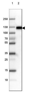
- Western Blot: LAMP-1/CD107a Antibody (3916) [NBP2-59780] - Lane 1: Marker [kDa] 250, 130, 100, 70, 55, 35, 25, 15, 10 Lane 2: Human cell line RT-4
Supportive validation
- Submitted by
- Novus Biologicals (provider)
- Main image
- Experimental details
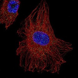
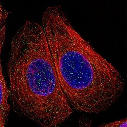
- Immunocytochemistry/Immunofluorescence: LAMP-1/CD107a Antibody (3916) [NBP2-59780] - Staining of U-251 cells showing specific staining of lysosomes in green. Microtubule and nuclear probes are visualized in red and blue, respectively (where available). Antibody staining is shown in green.

- Submitted by
- Novus Biologicals (provider)
- Main image
- Experimental details
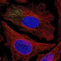
- Immunocytochemistry/Immunofluorescence: LAMP-1/CD107a Antibody (3916) [NBP2-59780] - Staining of HeLa cells showing specific staining of lysosomes in green. Microtubule and nuclear probes are visualized in red and blue, respectively (where available). Antibody staining is shown in green.

- Submitted by
- Novus Biologicals (provider)
- Main image
- Experimental details
- Immunocytochemistry/Immunofluorescence: LAMP-1/CD107a Antibody (3916) [NBP2-59780] - Staining of MCF7 cells showing specific staining of lysosomes in green. Microtubule and nuclear probes are visualized in red and blue, respectively (where available). Antibody staining is shown in green.

Supportive validation
- Submitted by
- Novus Biologicals (provider)
- Main image
- Experimental details
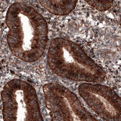
- Immunohistochemistry-Paraffin: LAMP-1/CD107a Antibody (3916) [NBP2-59780] - Staining of human endometrium shows granular cytoplasmic immunoreactivity in epithelial and stromal cells.

- Submitted by
- Novus Biologicals (provider)
- Main image

- Experimental details
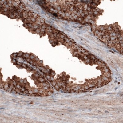
- Immunohistochemistry-Paraffin: LAMP-1/CD107a Antibody (3916) [NBP2-59780] - Staining of human prostate shows strong granular cytoplasmic positivity in epithelial cells.
- Submitted by
- Novus Biologicals (provider)
- Main image

- Experimental details
- Immunohistochemistry-Paraffin: LAMP-1/CD107a Antibody (3916) [NBP2-59780] - Staining of human fallopian tube shows granular cytoplasmic positivity in epithelial cells.

- Submitted by
- Novus Biologicals (provider)
- Main image

- Experimental details
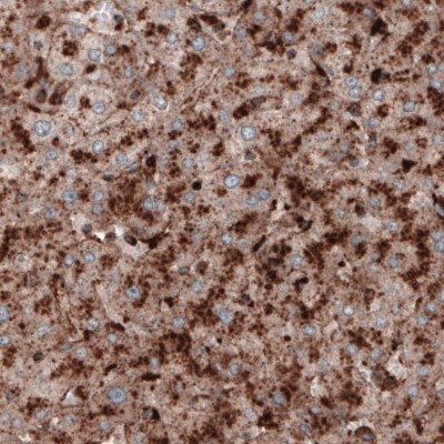
- Immunohistochemistry-Paraffin: LAMP-1/CD107a Antibody (3916) [NBP2-59780] - Staining of human liver shows strong granular cytoplasmic immunoreactivity in hepatocytes.
- Submitted by
- Novus Biologicals (provider)
- Main image
- Experimental details
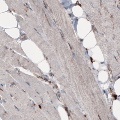
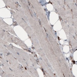
- Immunohistochemistry-Paraffin: LAMP-1/CD107a Antibody (3916) [NBP2-59780] - Staining of human skeletal muscle shows only very low cytoplasmic immunoreactivity.