Explore
 Validate
 Learn
Learn Western blot
 Immunocytochemistry
 Immunoprecipitation
 Immunohistochemistry
Antibody data
- Antibody Data
- Antigen structure
- References [0]
- Comments [0]
- Validations
- Western blot [1]
- Immunocytochemistry [3]
Submit
Validation data
Reference
Comment
Report error
- Product number
- LS-C783540 - Provider product page

- Provider
- LSBio
- Product name
- LAMP1 / CD107a Antibody (clone H4A3) LS-C783540
- Antibody type
- Monoclonal
- Description
- Purified by affinity chromatography.
- Reactivity
- Human, Porcine
- Host
- Mouse
- Isotype
- IgG
- Antibody clone number
- H4A3
- Storage
- Store at 2°C to 8°C.
No comments: Submit comment
Enhanced validation
- Submitted by
- LSBio (provider)
- Enhanced method
- Genetic validation
- Main image

- Experimental details
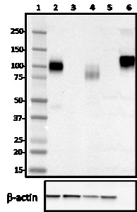
- Western blot of purified anti-LAMP-1 antibody (clone H4A3). Lane 1: Molecular weight marker; Lane 2: 20 µg of Hela cell lysate; Lane 3: 20 µg of NIH3T3 cell lysate; Lane 4: 20 µg of human brain lysate; Lane 5: 20 µg of mouse brain lysate; Lane 6: 10 ng of human recombinant LAMP-1. The blot was incubated with 1 µg/ml of the primary antibody overnight at 4°C, followed by incubation with HRP-labeled goat anti-mouse IgG. Anti-ß-actin antibody was used as the loading control. Enhanced chemiluminescence was used as the detection system.
Supportive validation
- Submitted by
- LSBio (provider)
- Enhanced method
- Genetic validation
- Main image

- Experimental details
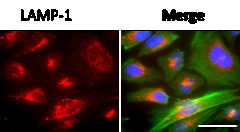
- ICC staining of purified anti-LAMP-1 antibody (clone H4A3) on Hela cells. The cells were fixed with 4% PFA, permeabilized with 0.1% Triton X-100, and blocked with 2% normal goat serum and 0.02% BSA. The cells were then stained with 5 µg/ml of the primary antibody overnight at 4°C, followed by incubation with Alexa Fluor® 594 goat anti-Mouse IgG for one hour at room temperature. Cells were counterstained with Phalloidin™ Green 488 and DAPI to visualize actin filaments and nuclei, respectively. The images were captured with a 60X objective. Scale bar: 50 µm.

- Submitted by
- LSBio (provider)
- Main image

- Experimental details
- ICC staining of purified anti-LAMP-1 antibody (clone H4A3) on Hela cells. The cells were fixed with 4% PFA, permeabilized with 0.1% Triton X-100, and blocked with 2% normal goat serum and 0.02% BSA. The cells were then stained with 5 µg/ml of the primary antibody overnight at 4°C, followed by incubation with Alexa Fluor® 594 goat anti-Mouse IgG for one hour at room temperature. Cells were counterstained with Phalloidin™ Green 488 and DAPI to visualize actin filaments and nuclei, respectively. The images were captured with a 60X objective. Scale bar: 50 µm.