Explore
 Validate
 Learn
LearnMA5-29385
antibody from Invitrogen Antibodies
Targeting: LAMP1
CD107a
 Western blot
 ELISA
ELISA Immunocytochemistry Immunoprecipitation Immunohistochemistry Flow cytometry Other assay
Antibody data
- Antibody Data
- Antigen structure
- References [1]
- Comments [0]
- Validations
- Immunocytochemistry [5]
- Immunoprecipitation [1]
- Immunohistochemistry [5]
- Flow cytometry [2]
- Other assay [2]
Submit
Validation data
Reference
Comment
Report error
- Product number
- MA5-29385 - Provider product page

- Provider
- Invitrogen Antibodies
- Product name
- LAMP1 Recombinant Rabbit Monoclonal Antibody (107)
- Antibody type
- Monoclonal
- Antigen
- Recombinant full-length protein
- Description
- This product is preservative free. It is recommended to add sodium azide to avoid contamination (final concentration 0.05%-0.1%). Recombinant rabbit monoclonal antibodies are produced using in vitro expression systems. The expression systems are developed by cloning in the specific antibody DNA sequences from immunoreactive rabbits. Then, individual clones are screened to select the best candidates for production. The advantages of using recombinant rabbit monoclonal antibodies include: better specificity and sensitivity, lot-to-lot consistency, animal origin-free formulations, and broader immunoreactivity to diverse targets due to larger rabbit immune repertoire. This antibody has specificity for Human LAMP1/CD107a.
- Reactivity
- Human, Rat
- Host
- Rabbit
- Isotype
- IgG
- Antibody clone number
- 107
- Vial size
- 100 µL
- Concentration
- 1 mg/mL
- Storage
- Store at 4°C short term. For long term storage, store at -20°C, avoiding freeze/thaw cycles.
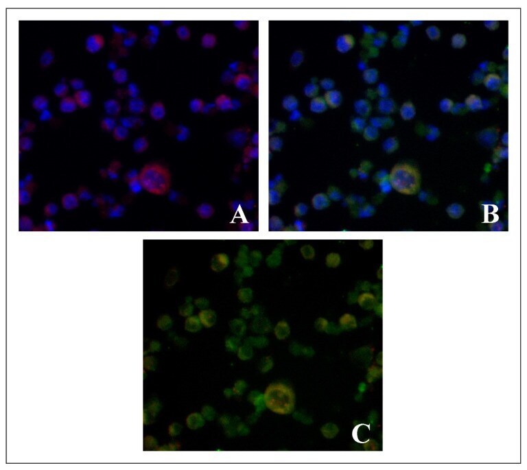
Submitted references Application of meso-CF(3)-Fluorophore BODIPY with Phenyl and Pyrazolyl Substituents for Lifetime Visualization of Lysosomes.
Trukhan IS, Tomilin DN, Dremina NN, Sobenina LN, Shurygin MG, Petrushenko KB, Petrushenko IK, Trofimov BA, Shurygina IA
Molecules (Basel, Switzerland) 2022 Aug 7;27(15)
Molecules (Basel, Switzerland) 2022 Aug 7;27(15)
No comments: Submit comment
Supportive validation
- Submitted by
- Invitrogen Antibodies (provider)
- Main image
- Experimental details
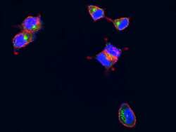
- Confocal immunofluorescence analysis of Human LAMP1 in MCF7 cells. Cells were fixed with 4% PFA, permeabilzed with 1% Triton X-100 in PBS, blocked with 10% serum, and incubated with LAMP1 Recombinant Rabbit Monoclonal Antibody (107) (Product # MA5-29385, 1:300). Then cells were stained with the Alexa Fluor® 488-conjugated Goat Anti-rabbit IgG secondary antibody, counterstained with Alexa Fluor® 546-conjugated phallotoxins (red) and DAPI (blue). Positive staining was localized to lysosome membrane.

- Submitted by
- Invitrogen Antibodies (provider)
- Main image

- Experimental details
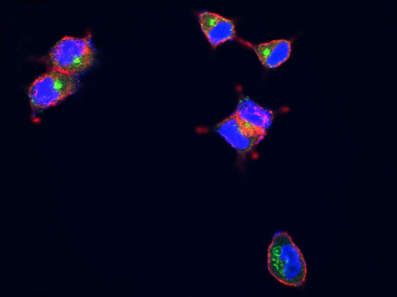
- Confocal immunofluorescence analysis of Human LAMP1 in MCF7 cells. Cells were fixed with 4% PFA, permeabilzed with 1% Triton X-100 in PBS, blocked with 10% serum, and incubated with LAMP1 Recombinant Rabbit Monoclonal Antibody (107) (Product # MA5-29385, 1:300). Then cells were stained with the Alexa Fluor® 488-conjugated Goat Anti-rabbit IgG secondary antibody, counterstained with Alexa Fluor® 546-conjugated phallotoxins (red) and DAPI (blue). Positive staining was localized to lysosome membrane.
- Submitted by
- Invitrogen Antibodies (provider)
- Main image

- Experimental details
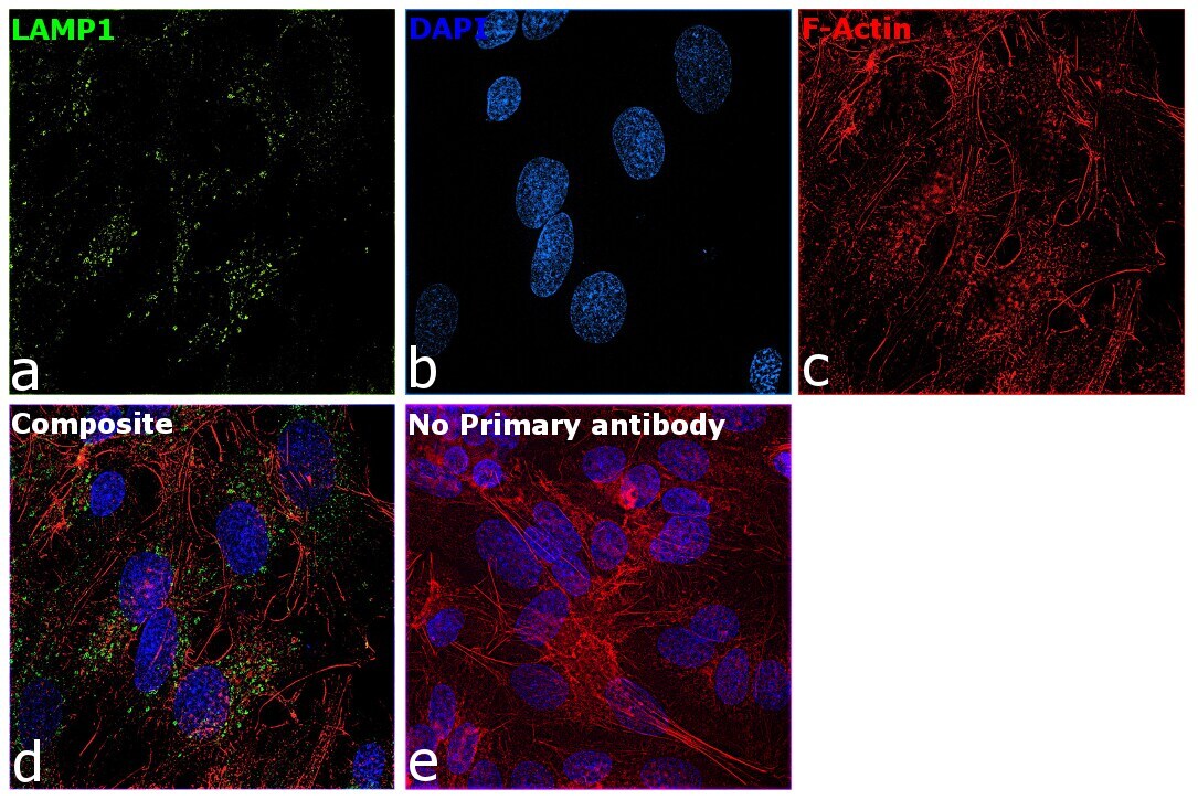
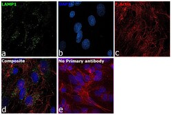
- Immunofluorescence analysis of LAMP1 (Lysosome-associated membrane glycoprotein 1) was performed using 80% confluent log phase Hep G2 cells. The cells were fixed with 4% paraformaldehyde for 10 minutes, permeabilized with 0.1% Triton™ X-100 for 10 minutes, and blocked with 2% BSA for 45 minutes at room temperature. The cells were labeled with LAMP1 Recombinant Rabbit Monoclonal Antibody (107) (Product # MA5-29385) at 1:200 in 0.1% BSA, incubated at 4 degree celsius overnight and then labeled with Donkey anti-Rabbit IgG (H+L) Highly Cross-Adsorbed Secondary Antibody, Alexa Fluor Plus 488 (Product # A32790), (1:2500 dilution), for 45 minutes at room temperature (Panel a: Green). Nuclei (Panel b:Blue) were stained with ProLong™ Diamond Antifade Mountant with DAPI (Product # P36962). F-actin (Panel c: Red) was stained with Rhodamine Phalloidin (Product # R415, 1:300). Panel d represents the merged image showing endosome and lysosome-like staining for LAMP1. Panel e represents control Hep G2 cells with no primary antibody to assess background. The images were captured at 60X magnification.
- Submitted by
- Invitrogen Antibodies (provider)
- Main image

- Experimental details
- Confocal immunofluorescence analysis of Human LAMP1 in MCF7 cells. Cells were fixed with 4% PFA, permeabilzed with 1% Triton X-100 in PBS, blocked with 10% serum, and incubated with LAMP1 Recombinant Rabbit Monoclonal Antibody (107) (Product # MA5-29385, 1:300). Then cells were stained with the Alexa Fluor® 488-conjugated Goat Anti-rabbit IgG secondary antibody, counterstained with Alexa Fluor® 546-conjugated phallotoxins (red) and DAPI (blue). Positive staining was localized to lysosome membrane.

- Submitted by
- Invitrogen Antibodies (provider)
- Main image
- Experimental details
- Immunofluorescence analysis of LAMP1 (Lysosome-associated membrane glycoprotein 1) was performed using 80% confluent log phase Hep G2 cells. The cells were fixed with 4% paraformaldehyde for 10 minutes, permeabilized with 0.1% Triton™ X-100 for 10 minutes, and blocked with 2% BSA for 45 minutes at room temperature. The cells were labeled with LAMP1 Recombinant Rabbit Monoclonal Antibody (107) (Product # MA5-29385) at 1:200 in 0.1% BSA, incubated at 4 degree celsius overnight and then labeled with Donkey anti-Rabbit IgG (H+L) Highly Cross-Adsorbed Secondary Antibody, Alexa Fluor Plus 488 (Product # A32790), (1:2500 dilution), for 45 minutes at room temperature (Panel a: Green). Nuclei (Panel b:Blue) were stained with ProLong™ Diamond Antifade Mountant with DAPI (Product # P36962). F-actin (Panel c: Red) was stained with Rhodamine Phalloidin (Product # R415, 1:300). Panel d represents the merged image showing endosome and lysosome-like staining for LAMP1. Panel e represents control Hep G2 cells with no primary antibody to assess background. The images were captured at 60X magnification.

Supportive validation
- Submitted by
- Invitrogen Antibodies (provider)
- Main image
- Experimental details
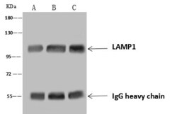
- Immunoprecipitation analysis of LAMP1 in Lane A: 0.5 mg Jurkat whole cell lysate, Lane B: 0.5 mg Hela whole cell lysate, Lane C: 0.5 mg Daudi whole cell lysate using 4 μL LAMP1 Recombinant Monoclonal (Product # MA5-29385) and 60 μg of Immunomagnetic beads Protein A/G. Primary antibody: LAMP1 Recombinant Monoclonal Antibody at a 1:100 dilution. Secondary antibody: Goat Anti-Rabbit IgG (H+L), HRP at 1:10,000 dilution. Developed using the ECL technique. Performed under reducing conditions. Predicted band size: 45 kDa. Observed band size : 120 kDa.

Supportive validation
- Submitted by
- Invitrogen Antibodies (provider)
- Main image
- Experimental details
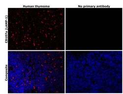
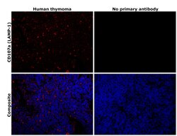
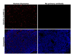
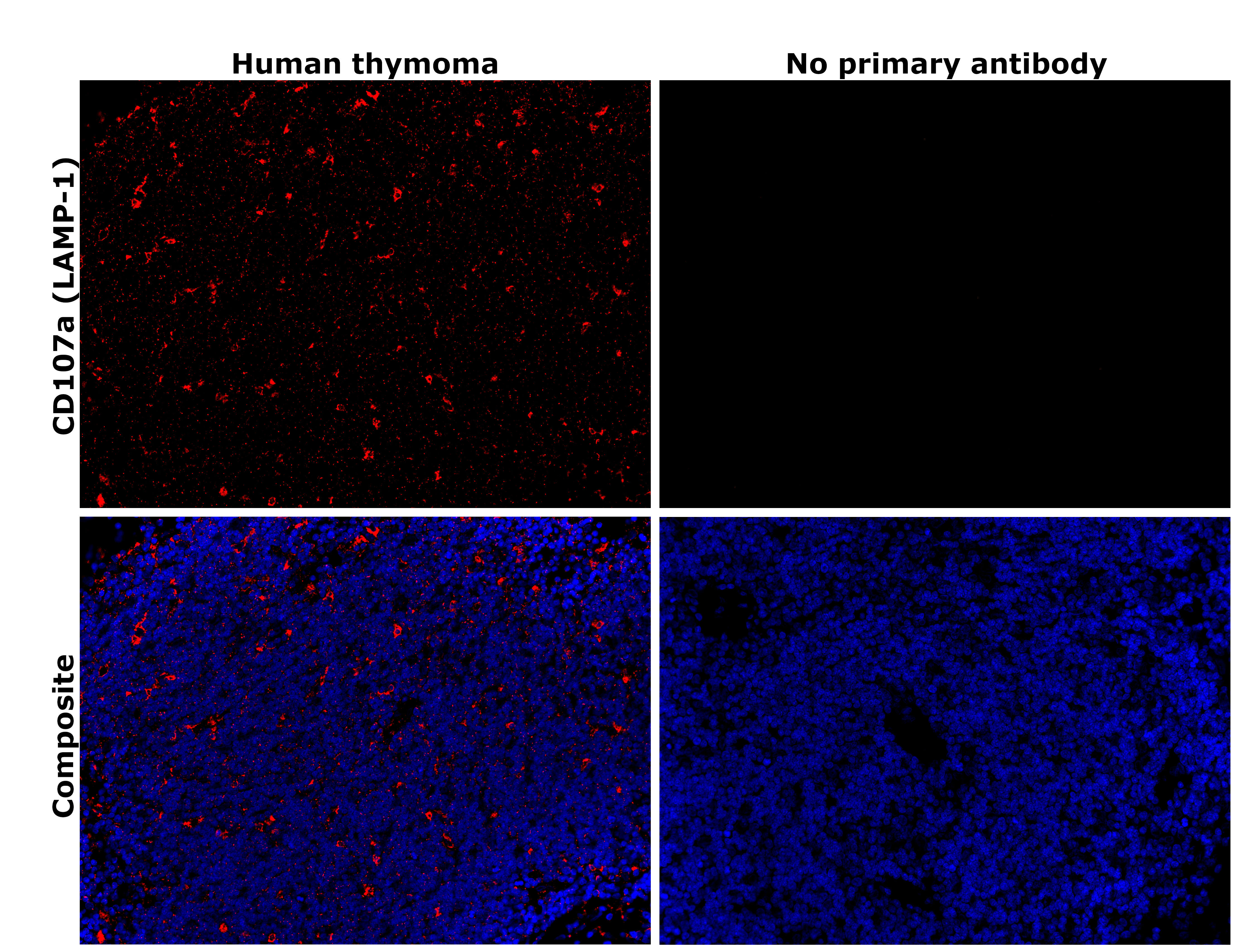
- Immunohistochemical analysis of CD107a (LAMP-1) was performed using formalin-fixed paraffin-embedded human cancer (thymoma) tissue sections. To expose the target protein, heat-induced epitope retrieval was performed on de-paraffinized sections using eBioscience™ IHC Antigen Retrieval Solution - High pH (10X) (Product # 00-4956-58) diluted to 1X solution in water in a decloaking chamber at 110 degree Celsius for 15 minutes. Following antigen retrieval, the sections were blocked with 3% H2O2 for 1 hour at room temperature followed by 2% normal goat serum in 1X PBS for 45 minutes at room temperature and then probed with or without LAMP1 Recombinant Rabbit Monoclonal Antibody (107) (Product # MA5-29385) at 1:500 dilution in 0.1% normal goat serum overnight at 4 degree Celsius in a humidified chamber. Detection was performed using Alexa Fluor™ 594 Tyramide SuperBoost™ Kit, goat anti-rabbit IgG (Product # B40925). Nuclei were stained with DAPI (Product # D1306) and the sections were mounted using ProLong™ Glass Antifade Mountant (Product # P36984). The images were captured on EVOS™ M7000 Imaging System (Product # AMF7000) at 20X magnification and externally deconvoluted.
- Submitted by
- Invitrogen Antibodies (provider)
- Main image

- Experimental details
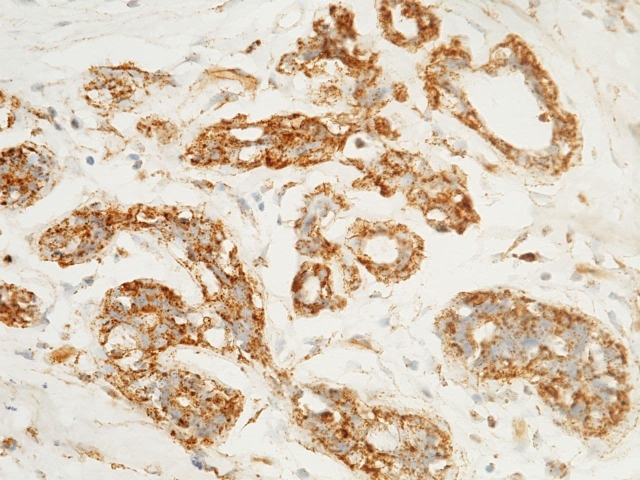
- Immunohistochemical staining of human LAMP1 in human breast carcinoma with LAMP1 Recombinant Rabbit Monoclonal Antibody (107) (Product # MA5-29385, 1:1,000, formalin-fixed paraffin embedded sections).
- Submitted by
- Invitrogen Antibodies (provider)
- Main image
- Experimental details
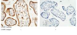
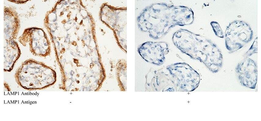
- Immunohistochemical staining of human LAMP1 in human placenta with LAMP1 Recombinant Rabbit Monoclonal Antibody (107) (Product # MA5-29385, 1:1,000, formalin-fixed paraffin embedded sections). Left panel: tissue incubated with primary antibody. Right panel: tissue incubated with mixture of primary antibody and antigen (recombinant protein).
- Submitted by
- Invitrogen Antibodies (provider)
- Main image
- Experimental details
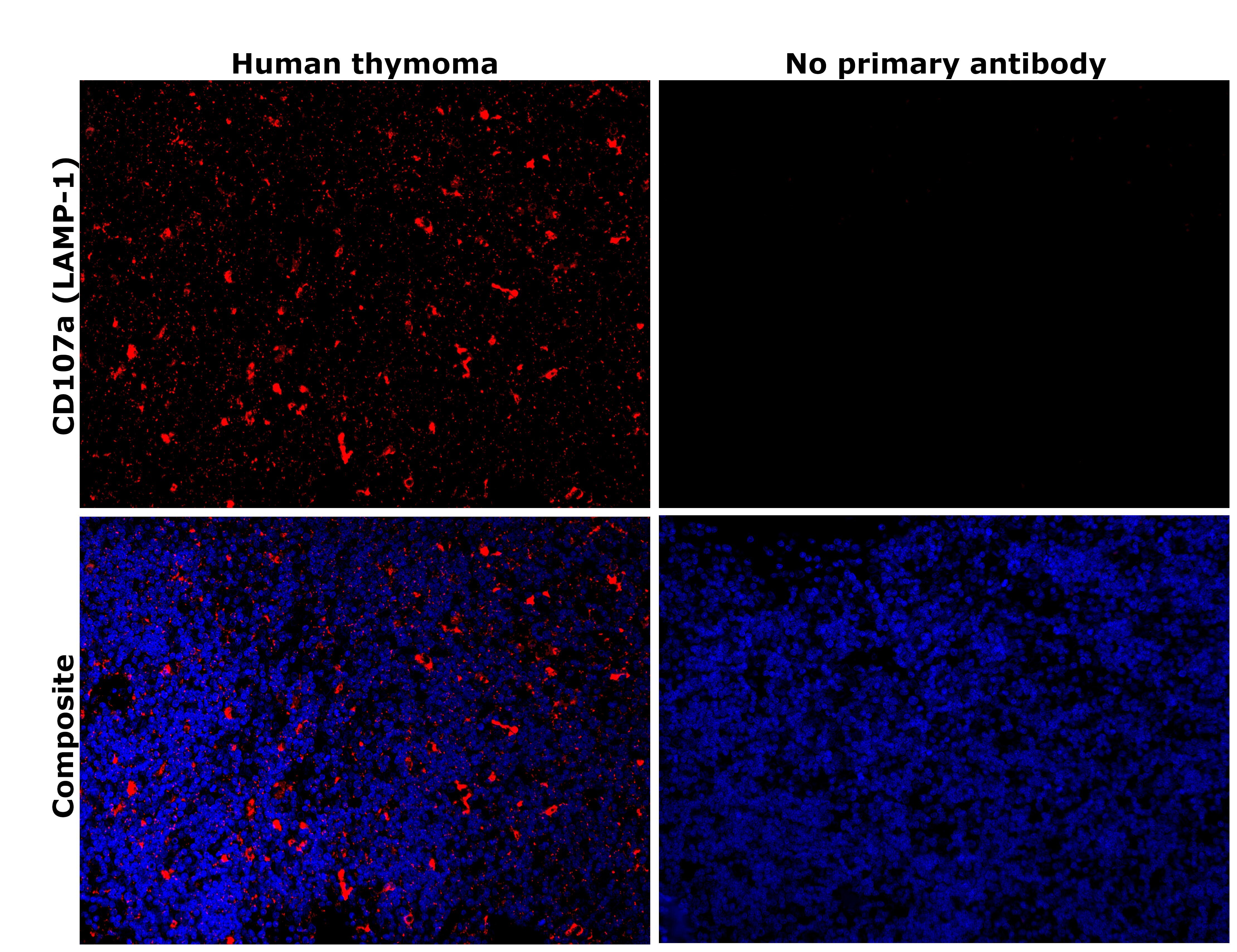
- Immunohistochemical analysis of CD107a (LAMP-1) was performed using formalin-fixed paraffin-embedded human cancer (thymoma) tissue sections. To expose the target protein, heat-induced epitope retrieval was performed on de-paraffinized sections using eBioscience™ IHC Antigen Retrieval Solution - Low pH (10X) (Product # 00-4955-58) diluted to 1X solution in water in a decloaking chamber at 110 degree Celsius for 15 minutes. Following antigen retrieval, the sections were blocked with 3% H2O2 for 1 hour at room temperature followed by 2% normal goat serum in 1X PBS for 45 minutes at room temperature and then probed with or without LAMP1 Recombinant Rabbit Monoclonal Antibody (107) (Product # MA5-29385) at 1:500 dilution in 0.1% normal goat serum overnight at 4 degree Celsius in a humidified chamber. Detection was performed using Alexa Fluor™ 594 Tyramide SuperBoost™ Kit, goat anti-rabbit IgG (Product # B40925). Nuclei were stained with DAPI (Product # D1306) and the sections were mounted using ProLong™ Glass Antifade Mountant (Product # P36984). The images were captured on EVOS™ M7000 Imaging System (Product # AMF7000) at 20X magnification and externally deconvoluted.

- Submitted by
- Invitrogen Antibodies (provider)
- Main image
- Experimental details
- Immunohistochemical analysis of CD107a (LAMP-1) was performed using formalin-fixed paraffin-embedded human cancer (thymoma) tissue sections. To expose the target protein, heat-induced epitope retrieval was performed on de-paraffinized sections using eBioscience™ IHC Antigen Retrieval Solution - High pH (10X) (Product # 00-4956-58) diluted to 1X solution in water in a decloaking chamber at 110 degree Celsius for 15 minutes. Following antigen retrieval, the sections were blocked with 2% normal goat serum in 1X PBS for 45 minutes at room temperature and then probed with or without LAMP1 Recombinant Rabbit Monoclonal Antibody (107) (Product # MA5-29385) at 1:200 dilution in 0.1% normal goat serum overnight at 4 degree Celsius in a humidified chamber. Detection was performed using Goat anti-Rabbit IgG (H+L) Highly Cross-Adsorbed Secondary Antibody, Alexa Fluor™ Plus 647 (Product # A32733) at a dilution of 1:2,000 in 0.1% normal goat serum for 45 minutes at room temperature. ReadyProbes™ Tissue Autofluorescence Quenching Kit (Product # R37630) was used to quench autofluorescence from the tissues. Nuclei were stained with DAPI (Product # D1306) and the sections were mounted using ProLong™ Glass Antifade Mountant (Product # P36984). The images were captured on EVOS™ M7000 Imaging System (Product # AMF7000) at 20X magnification and externally deconvoluted.
Supportive validation
- Submitted by
- Invitrogen Antibodies (provider)
- Main image
- Experimental details
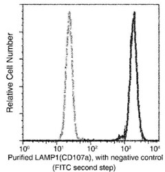
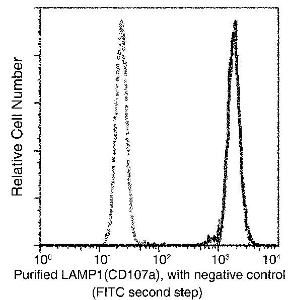
- Flow cytometric analysis of Human LAMP1 on Jurkat cells. Cells were treated according to manufacturer’s manual, stained with LAMP1 Recombinant Rabbit Monoclonal Antibody (107) (Product # MA5-29385), then a FITC-conjugated Secondary antibody. The histograms were derived from gated events with the forward and side light-scatter characteristics of intact cells.

- Submitted by
- Invitrogen Antibodies (provider)
- Main image

- Experimental details
- Flow cytometric analysis of Human LAMP1 on Jurkat cells. Cells were treated according to manufacturer’s manual, stained with LAMP1 Recombinant Rabbit Monoclonal Antibody (107) (Product # MA5-29385), then a FITC-conjugated Secondary antibody. The histograms were derived from gated events with the forward and side light-scatter characteristics of intact cells.
Supportive validation
- Submitted by
- Invitrogen Antibodies (provider)
- Main image

- Experimental details
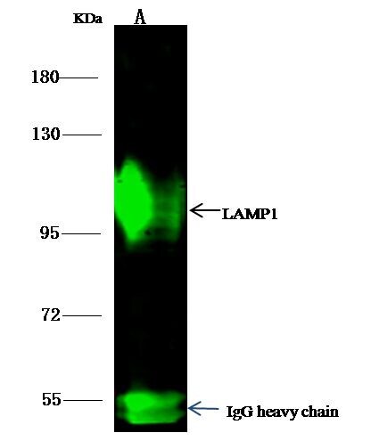
- LAMP1 Immunoprecipitation using: Lane A: 0.5 mg Jurkat Whole Cell Lysate 1 µL with LAMP1 Recombinant Rabbit Monoclonal Antibody (107) (Product # MA5-29385) and 15 µL of 50 % Protein G agarose. Primary antibody: LAMP1 Recombinant Rabbit Monoclonal Antibody (107), at 1:500 dilution. Secondary antibody: Dylight 800-labeled antibody to rabbit IgG (H+L), at 1:5,000 dilution. Developed using the Odyssey technique. Performed under reducing conditions. Predicted band size: 45 kDa. Observed band size: 113 kDa.
- Submitted by
- Invitrogen Antibodies (provider)
- Main image

- Experimental details
- Fluorescent microscopic images showing colocalization of lysosomal markers LAMP1 and BODIPY 1 in Ehrlich ascitic carcinoma cells: ( A )--BODIPY 1 (red) and nuclei (blue), ( B )--LAMP1 (green) and nuclei (blue), ( C )--overlapping BODIPY 1 and LAMP1 fluorescent staining. Magnification 40x.