 Explore
 Validate
 Learn
Learn Western blot
Antibody data
- Antibody Data
- Antigen structure
- References [1]
- Comments [0]
- Validations
- Western blot [1]
- Immunocytochemistry [1]
Submit
Validation data
Reference
Comment
Report error
- Product number
- AF4800 - Provider product page

- Provider
- R&D Systems
- Product name
- Human LAMP-1/CD107a Lumenal Domain Antibody
- Antibody type
- Polyclonal
- Description
- Antigen Affinity-purified. Detects human LAMP1/CD107a Lumenal Domain in direct ELISAs and Western blots. In direct ELISAs, less than 1% cross-reactivity with recombinant mouse LAMP1 is observed.
- Reactivity
- Human
- Host
- Sheep
- Conjugate
- Unconjugated
- Antigen sequence
P11279- Isotype
- IgG
- Vial size
- 100 ug
- Concentration
- LYOPH
- Storage
- Use a manual defrost freezer and avoid repeated freeze-thaw cycles. 12 months from date of receipt, -20 to -70 °C as supplied. 1 month, 2 to 8 °C under sterile conditions after reconstitution. 6 months, -20 to -70 °C under sterile conditions after reconstitution.
Submitted references Cellular trafficking of lipoteichoic acid and Toll-like receptor 2 in relation to signaling: role of CD14 and CD36.
Nilsen NJ, Deininger S, Nonstad U, Skjeldal F, Husebye H, Rodionov D, von Aulock S, Hartung T, Lien E, Bakke O, Espevik T
Journal of leukocyte biology 2008 Jul;84(1):280-91
Journal of leukocyte biology 2008 Jul;84(1):280-91
No comments: Submit comment
Supportive validation
- Submitted by
- R&D Systems (provider)
- Main image

- Experimental details
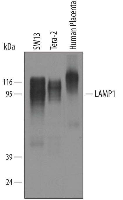
- Detection of Human LAMP1/CD107a by Western Blot. Western blot shows lysates of SW13 human adrenal cortex adenocarcinoma cell line, Tera-2 human embryonic lung carcinoma cell line, and human placenta tissue. PVDF membrane was probed with 1 µg/mL of Sheep Anti-Human LAMP1/CD107a Lumenal Domain Antigen Affinity-purified Polyclonal Antibody (Catalog # AF4800) followed by HRP-conjugated Anti-Sheep IgG Secondary Antibody (Catalog # HAF016). A specific band was detected for LAMP1/CD107a at approximately 90 - 120 kDa (as indicated). This experiment was conducted under reducing conditions and using Immunoblot Buffer Group 8.
Supportive validation
- Submitted by
- R&D Systems (provider)
- Main image

- Experimental details
- LAMP1/CD107a in THP-1 Human Cell Line. LAMP1/CD107a was detected in immersion fixed THP-1 human acute monocytic leukemia cell line using 10 µg/mL Sheep Anti-Human LAMP1/CD107a Lumenal Domain Antigen Affinity-purified Polyclonal Antibody (Catalog # AF4800) for 3 hours at room temperature. Cells were stained with the NorthernLights™ 557-conjugated Anti-Sheep IgG Secondary Antibody (red; Catalog # NL010) and counterstained with DAPI (blue). View our protocol for Fluorescent ICC Staining of Cells on Coverslips.
