Explore
 Validate
 Learn
Learn Western blot
Antibody data
- Antibody Data
- Antigen structure
- References [1]
- Comments [0]
- Validations
- Western blot [2]
- Immunocytochemistry [8]
- Other assay [1]
Submit
Validation data
Reference
Comment
Report error
- Product number
- PA1-10022 - Provider product page

- Provider
- Invitrogen Antibodies
- Product name
- Coronin 1A Polyclonal Antibody
- Antibody type
- Polyclonal
- Antigen
- Synthetic peptide
- Description
- PA1-10022 was made against the C-terminal peptide of human coronin 1a chemically coupled to keyhole limpet hemocyanin carrier protein. PA1-10022 is also known to work on many mammalian species both on western blots and on cells grown in tissue culture.
- Reactivity
- Human, Mouse, Rat, Bovine, Porcine
- Host
- Rabbit
- Isotype
- IgG
- Vial size
- 100 μL
- Concentration
- Conc. Not Determined
- Storage
- Store at 4°C short term. For long term storage, store at -20°C, avoiding freeze/thaw cycles.
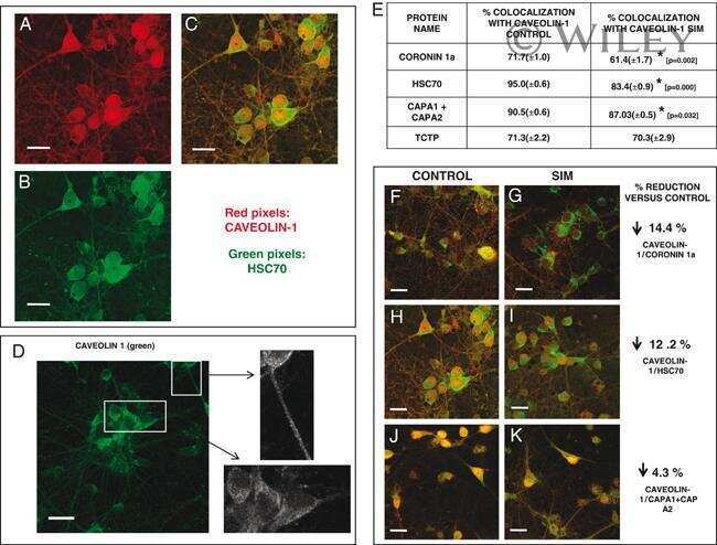
Submitted references The effect of simvastatin on the proteome of detergent-resistant membrane domains: decreases of specific proteins previously related to cytoskeleton regulation, calcium homeostasis and cell fate.
Ponce J, Brea D, Carrascal M, Guirao V, Degregorio-Rocasolano N, Sobrino T, Castillo J, Dávalos A, Gasull T
Proteomics 2010 May;10(10):1954-65
Proteomics 2010 May;10(10):1954-65
No comments: Submit comment
Supportive validation
- Submitted by
- Invitrogen Antibodies (provider)
- Main image

- Experimental details
- Western blot of coronin 1a in tissue lysates using rabbit pAb to coronin 1a: protein standard (Lane 1), mouse brain (Lane 2), rat brain (Lane 3), cow cerebellum (Lane 4), cow cortex (Lane 5), and pig spinal cord (Lane 6). The strong single band about 55 kDa corresponds to the coronin 1a polyclonal antibody (Product # PA1-10022, dilution 1:5,000).

- Submitted by
- Invitrogen Antibodies (provider)
- Main image

- Experimental details
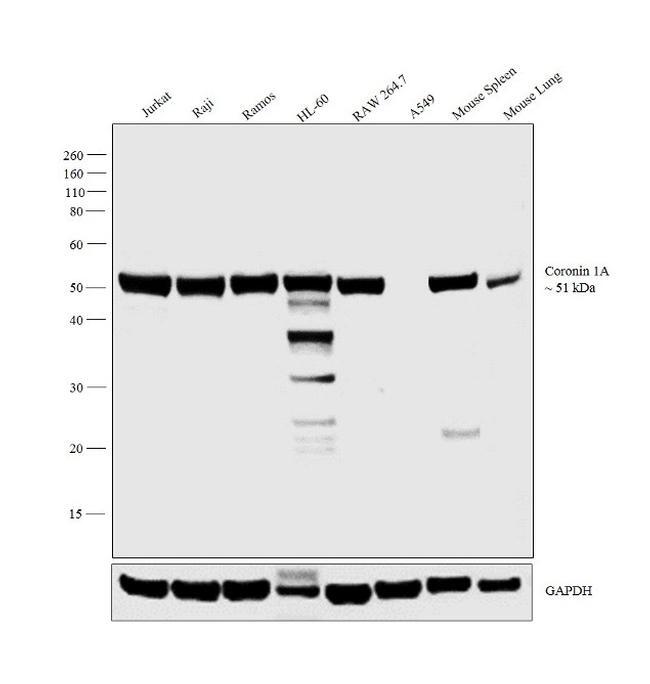
- Western blot analysis was performed on membrane enriched extracts (30 µg lysate) of Jurkat (Lane 1), Raji (Lane 2), Ramos (Lane 3), HL-60 (Lane 4), RAW 264.7 (Lane 5), A549 (Lane 6), tissue extracts of Mouse Spleen (Lane 7) and Mouse Lung (Lane 8). The blot was probed with Anti-Coronin 1A Polyclonal Antibody (Product # PA1-10022, 1:4,000 dilution) and detected by chemiluminescence using Goat anti-Rabbit IgG (Heavy Chain) Superclonal™ Secondary Antibody, HRP conjugate (Product # A27036, 0.25 µg/mL, 1:4,000 dilution). A 51 kDa band corresponding to Coronin 1A was observed across the cell lines and tissues tested except in A549 which is reported to be negative for Coronin 1A expression.
Supportive validation
- Submitted by
- Invitrogen Antibodies (provider)
- Main image

- Experimental details
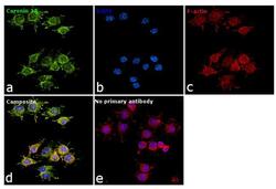
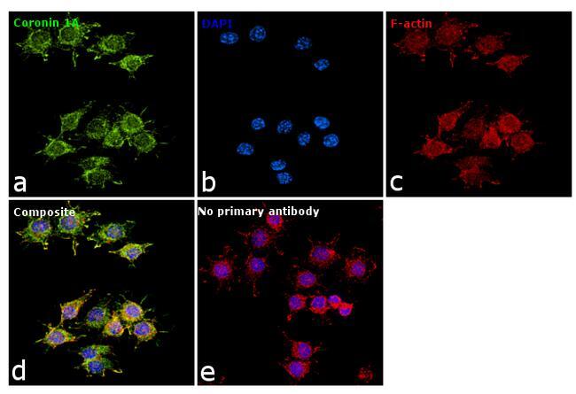
- Immunofluorescence analysis of Coronin 1A was performed using 70% confluent log phase RAW 264.7 cells. The cells were fixed with 4% paraformaldehyde for 10 minutes, permeabilized with 0.1% Triton™ X-100 for 15 minutes, and blocked with 1% BSA for 1 hour at room temperature. The cells were labeled with Coronin 1A Polyclonal Antibody (Product # PA1-10022) at 1:100 dilution in 0.1% BSA, incubated at 4 degree Celsius overnight and then labeled with Goat anti-Rabbit IgG (H+L) Superclonal™ Secondary Antibody, Alexa Fluor® 488 conjugate (Product # A27034) at a dilution of 1:2000 for 45 minutes at room temperature (Panel a: green). Nuclei (Panel b: blue) were stained with ProLong™ Diamond Antifade Mountant with DAPI (Product # P36962). F-actin (Panel c: red) was stained with Rhodamine Phalloidin (Product # R415). Panel d represents the merged image showing cytoskeletal localization. Panel e represents control cells with no primary antibody to assess background. The images were captured at 60X magnification.

- Submitted by
- Invitrogen Antibodies (provider)
- Main image

- Experimental details
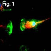
- Immunofluorescent analysis of Coronin 1A using a polyclonal antibody (Product # PA1-10022).

- Submitted by
- Invitrogen Antibodies (provider)
- Main image
- Experimental details
- Immunofluorescent analysis of Coronin 1A using a polyclonal antibody (Product # PA1-10022).

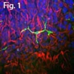
- Submitted by
- Invitrogen Antibodies (provider)
- Main image
- Experimental details
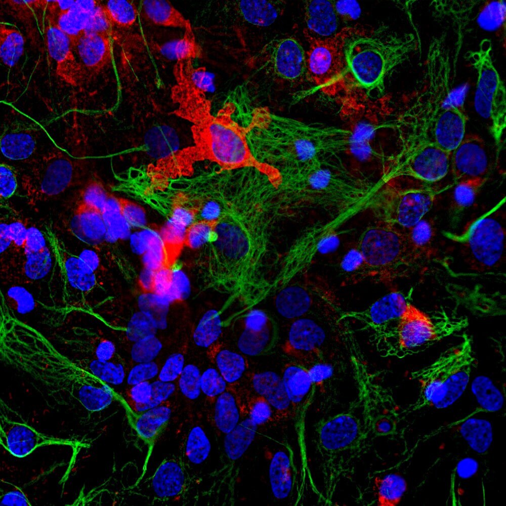
- Immunofluorescent analysis of cortical neuron-glial cell culture from E20 rat stained with rabbit pAb to coronin 1a (Product # PA1-10022, diluted 1:1,000) in red, and costained with mouse mAb to GFAP (diluted 1:1,000) in green. The blue is Hoechst staining of nuclear DNA. The coronin 1a antibody labels protein expressed in the cytoplasm of microglia cells, while GFAP antibody stains intermediate filaments in astrocytic cells.
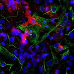
- Submitted by
- Invitrogen Antibodies (provider)
- Main image

- Experimental details
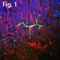
- Immunofluorescent analysis of Coronin 1A using a polyclonal antibody (Product # PA1-10022).
- Submitted by
- Invitrogen Antibodies (provider)
- Main image

- Experimental details
- Immunofluorescent analysis of Coronin 1A using a polyclonal antibody (Product # PA1-10022).
- Submitted by
- Invitrogen Antibodies (provider)
- Main image
- Experimental details
- Immunofluorescence analysis of Coronin 1A was performed using 70% confluent log phase RAW 264.7 cells. The cells were fixed with 4% paraformaldehyde for 10 minutes, permeabilized with 0.1% Triton™ X-100 for 15 minutes, and blocked with 1% BSA for 1 hour at room temperature. The cells were labeled with Coronin 1A Polyclonal Antibody (Product # PA1-10022) at 1:100 dilution in 0.1% BSA, incubated at 4 degree Celsius overnight and then labeled with Goat anti-Rabbit IgG (Heavy Chain) Superclonal™ Secondary Antibody, Alexa Fluor® 488 conjugate (Product # A27034) at a dilution of 1:2000 for 45 minutes at room temperature (Panel a: green). Nuclei (Panel b: blue) were stained with ProLong™ Diamond Antifade Mountant with DAPI (Product # P36962). F-actin (Panel c: red) was stained with Rhodamine Phalloidin (Product # R415). Panel d represents the merged image showing cytoskeletal localization. Panel e represents control cells with no primary antibody to assess background. The images were captured at 60X magnification.
Supportive validation
- Submitted by
- Invitrogen Antibodies (provider)
- Main image
- Experimental details
- NULL