 Explore
 Validate
 Learn
Learn Western blot
 Immunocytochemistry
Antibody data
- Antibody Data
- Antigen structure
- References [0]
- Comments [0]
- Validations
- Immunocytochemistry [2]
- Immunoprecipitation [1]
- Chromatin Immunoprecipitation [4]
- Other assay [2]
Submit
Validation data
Reference
Comment
Report error
- Product number
- 702490 - Provider product page

- Provider
- Invitrogen Antibodies
- Product name
- SUZ12 Recombinant Rabbit Monoclonal Antibody (7H26L21)
- Antibody type
- Monoclonal
- Antigen
- Other
- Description
- This antibody is predicted to react with Monkey, Bovine, Pig, Mouse Recombinant rabbit monoclonal antibodies are produced using in vitro expression systems. The expression systems are developed by cloning in the specific antibody DNA sequences from immunoreactive rabbits. Then, individual clones are screened to select the best candidates for production. The advantages of using recombinant rabbit monoclonal antibodies include: better specificity and sensitivity, lot-to-lot consistency, animal origin-free formulations, and broader immunoreactivity to diverse targets due to larger rabbit immune repertoire.
- Reactivity
- Human
- Host
- Rabbit
- Isotype
- IgG
- Antibody clone number
- 7H26L21
- Vial size
- 100 μg
- Concentration
- 0.5 mg/mL
- Storage
- Store at 4°C short term. For long term storage, store at -20°C, avoiding freeze/thaw cycles.
No comments: Submit comment
Supportive validation
- Submitted by
- Invitrogen Antibodies (provider)
- Main image
- Experimental details
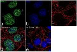
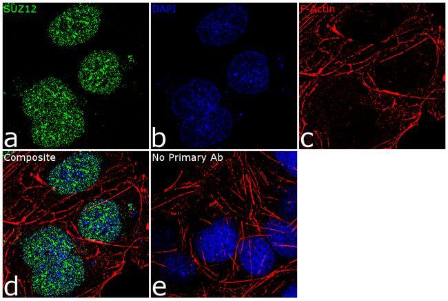
- For immunofluorescence analysis, NTERA-2 cells were fixed and permeabilized for detection of endogenous SUZ12 using Anti- SUZ12 Recombinant Rabbit Monoclonal Antibody (Product # 702490, 2 µg/mL) and labeled with Goat anti-Rabbit IgG (H+L) Superclonal™ Secondary Antibody, Alexa Fluor® 488 conjugate (Product # A27034, 1:2000). Panel a) shows representative cells that were stained for detection and localization of SUZ12 protein (green), Panel b) is stained for nuclei (blue) using SlowFade® Gold Antifade Mountant with DAPI (Product # S36938). Panel c) represents cytoskeletal F-actin staining using Rhodamine Phalloidin (Product # R415, 1:300). Panel d) is a composite image of panels a, b and c clearly demonstrating nuclear localization of SUZ12. Panel e) represents control cells with no primary antibody to assess background. The images were captured at 60X magnification.

- Submitted by
- Invitrogen Antibodies (provider)
- Main image
- Experimental details
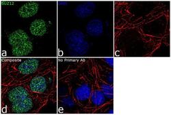
- For immunofluorescence analysis, NTERA-2 cells were fixed and permeabilized for detection of endogenous SUZ12 using Anti- SUZ12 Recombinant Rabbit Monoclonal Antibody (Product # 702490, 2 µg/mL) and labeled with Goat anti-Rabbit IgG (Heavy Chain) Superclonal™ Secondary Antibody, Alexa Fluor® 488 conjugate (Product # A27034, 1:2000). Panel a) shows representative cells that were stained for detection and localization of SUZ12 protein (green), Panel b) is stained for nuclei (blue) using SlowFade® Gold Antifade Mountant with DAPI (Product # S36938). Panel c) represents cytoskeletal F-actin staining using Rhodamine Phalloidin (Product # R415, 1:300). Panel d) is a composite image of panels a, b and c clearly demonstrating nuclear localization of SUZ12. Panel e) represents control cells with no primary antibody to assess background. The images were captured at 60X magnification.
Supportive validation
- Submitted by
- Invitrogen Antibodies (provider)
- Main image
- Experimental details
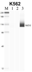
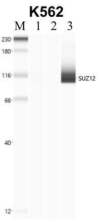
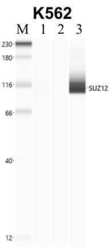
- Immunoprecipitation of SUZ12 was performed in K562 cells. Antigen-antibody complexes were formed by incubating approximately 500 µg whole cell lysate with 5 to 10 µL of recombinant monoclonal SUZ12 antibody (Product # 702490) rotating 60 min at RT. The immune complexes were captured on 625 µg of anti- rabbit coated Dynabeads (Product # 11204D) and washed extensively. They were then eluted and analyzed using the Simple Western system using the same antibody as used in immunoprecipitation at a dilution of 1:25, followed by a 1:100 dilution of secondary antibody. Lane 1 is the input, lane 2 no antibody IP and lane 3 is the target specific IP. Data courtesy of the Yeo lab as part of the ENCODE project.
Supportive validation
- Submitted by
- Invitrogen Antibodies (provider)
- Main image
- Experimental details
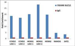
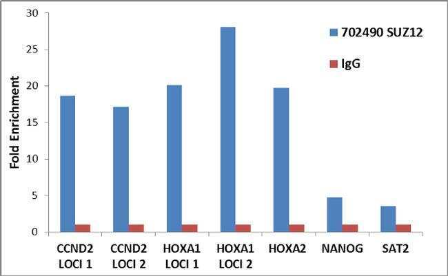
- Enrichment of endogenous SUZ12 protein at specific gene loci using Anti-SUZ12 Recombinant Rabbit Monoclonal Antibody: Chromatin Immunoprecipitation (ChIP) was performed using Anti-SUZ12 Recombinant Rabbit Monoclonal Antibody (Product # 702490, 5 µg) on sheared chromatin from 2 million Ntera2 cells using the MAGnify ChIP system kit (Product # 49-2024). Normal Rabbit IgG (1 µg) was used as a negative IP control. The purified DNA was analyzed by 7500 Fast qPCR system (Product # 4351106) with optimized PCR primer pairs for the different loci of the active CCND2, HOXA1, HOXA2 promoter region used as positive control target genes, and the region of the inactive NANOG promoter, SAT2 satellite repeat, used as negative control target gene. Data is presented as fold enrichment of the antibody signal versus the negative control IgG using the comparative CT method.
- Submitted by
- Invitrogen Antibodies (provider)
- Main image

- Experimental details
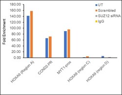
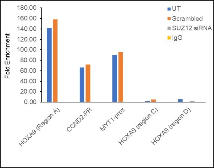
- Chromatin Immunoprecipitation (ChIP) assay of endogenous SUZ12 protein using SUZ12 Polyclonal Antibody: ChIP was performed using SUZ12 Polyclonal Antibody (Product # 702490, 5 µg) on sheared chromatin from HeLa cells using the MAGnify ChIP System kit (Product # 49-2024). Normal Rabbit IgG was used as a negative IP control. The purified DNA was analyzed by qPCR using primers binding to HOXA9 (Region A), CCND2-PR, MYT1-Prox as active binding regions and HOXA9 (region C) and HOXA9 (region D) as inactive binding regions. Data is presented as fold enrichment of the antibody signal versus the negative control IgG using the comparative CT method. PR: Promoter, Region A: -3.75 kb of HOXA9 promoter, Region B: +2 Kb of HOXA9 promoter, Region D: +5 kb of HOXA9 promoter, prox- proximal to MYT1 promoter.

- Submitted by
- Invitrogen Antibodies (provider)
- Main image

- Experimental details
- Enrichment of endogenous SUZ12 protein at specific gene loci using Anti-SUZ12 Recombinant Rabbit Monoclonal Antibody: Chromatin Immunoprecipitation (ChIP) was performed using Anti-SUZ12 Recombinant Rabbit Monoclonal Antibody (Product # 702490, 5 µg) on sheared chromatin from 2 million Ntera2 cells using the MAGnify ChIP system kit (Product # 49-2024). Normal Rabbit IgG (1 µg) was used as a negative IP control. The purified DNA was analyzed by 7500 Fast qPCR system (Product # 4351106) with optimized PCR primer pairs for the different loci of the active CCND2, HOXA1, HOXA2 promoter region used as positive control target genes, and the region of the inactive NANOG promoter, SAT2 satellite repeat, used as negative control target gene. Data is presented as fold enrichment of the antibody signal versus the negative control IgG using the comparative CT method.

- Submitted by
- Invitrogen Antibodies (provider)
- Main image
- Experimental details
- Chromatin Immunoprecipitation (ChIP) assay of endogenous SUZ12 protein using SUZ12 Polyclonal Antibody: ChIP was performed using SUZ12 Polyclonal Antibody (Product # 702490, 5 µg) on sheared chromatin from HeLa cells using the MAGnify ChIP System kit (Product # 49-2024). Normal Rabbit IgG was used as a negative IP control. The purified DNA was analyzed by qPCR using primers binding to HOXA9 (Region A), CCND2-PR, MYT1-Prox as active binding regions and HOXA9 (region C) and HOXA9 (region D) as inactive binding regions. Data is presented as fold enrichment of the antibody signal versus the negative control IgG using the comparative CT method. PR: Promoter, Region A: -3.75 kb of HOXA9 promoter, Region B: +2 Kb of HOXA9 promoter, Region D: +5 kb of HOXA9 promoter, prox- proximal to MYT1 promoter.
Supportive validation
- Submitted by
- Invitrogen Antibodies (provider)
- Main image
- Experimental details
- Immunoprecipitation of SUZ12 was performed in K562 cells. Antigen-antibody complexes were formed by incubating approximately 500 µg whole cell lysate with 5 to 10 µL of recombinant monoclonal SUZ12 antibody (Product # 702490) rotating 60 min at RT. The immune complexes were captured on 625 µg of anti- rabbit coated Dynabeads (Product # 11204D) and washed extensively. They were then eluted and analyzed using the Simple Western system using the same antibody as used in immunoprecipitation at a dilution of 1:25, followed by a 1:100 dilution of secondary antibody. Lane 1 is the input, lane 2 no antibody IP and lane 3 is the target specific IP. Data courtesy of the Yeo lab as part of the ENCODE project.

- Submitted by
- Invitrogen Antibodies (provider)
- Main image

- Experimental details
- Immunoprecipitation of SUZ12 was performed in K562 cells. Antigen-antibody complexes were formed by incubating approximately 500 µg whole cell lysate with 5 to 10 µL of recombinant monoclonal SUZ12 antibody (Product # 702490) rotating 60 min at RT. The immune complexes were captured on 625 µg of anti- rabbit coated Dynabeads (Product # 11204D) and washed extensively. They were then eluted and analyzed using the Simple Western system using the same antibody as used in immunoprecipitation at a dilution of 1:25, followed by a 1:100 dilution of secondary antibody. Lane 1 is the input, lane 2 no antibody IP and lane 3 is the target specific IP. Data courtesy of the Yeo lab as part of the ENCODE project.
