Explore
 Validate
 Learn
Learn Western blot
Antibody data
- Antibody Data
- Antigen structure
- References [3]
- Comments [0]
- Validations
- Western blot [1]
- Immunohistochemistry [1]
- Other assay [3]
Submit
Validation data
Reference
Comment
Report error
- Product number
- PA5-30989 - Provider product page

- Provider
- Invitrogen Antibodies
- Product name
- SERPINA12 Polyclonal Antibody
- Antibody type
- Polyclonal
- Antigen
- Recombinant full-length protein
- Description
- Recommended positive controls: 293T, A431, Jurkat, Raji. Store product as a concentrated solution. Centrifuge briefly prior to opening the vial.
- Reactivity
- Human
- Host
- Rabbit
- Isotype
- IgG
- Vial size
- 100 μL
- Concentration
- 0.57 mg/mL
- Storage
- Store at 4°C short term. For long term storage, store at -20°C, avoiding freeze/thaw cycles.
Submitted references Flutamide Alters the Expression of Chemerin, Apelin, and Vaspin and Their Respective Receptors in the Testes of Adult Rats.
Expression and Impact of Vaspin on In Vitro Oocyte Maturation through MAP3/1 and PRKAA1 Signalling Pathways.
Vaspin in the pig ovarian follicles: expression and regulation by different hormones.
Brzoskwinia M, Pardyak L, Rak A, Kaminska A, Hejmej A, Marek S, Kotula-Balak M, Bilinska B
International journal of molecular sciences 2020 Jun 22;21(12)
International journal of molecular sciences 2020 Jun 22;21(12)
Expression and Impact of Vaspin on In Vitro Oocyte Maturation through MAP3/1 and PRKAA1 Signalling Pathways.
Kurowska P, Mlyczyńska E, Estienne A, Barbe A, Rajska I, Soból K, Poniedziałek-Kempny K, Dupont J, Rak A
International journal of molecular sciences 2020 Dec 8;21(24)
International journal of molecular sciences 2020 Dec 8;21(24)
Vaspin in the pig ovarian follicles: expression and regulation by different hormones.
Kurowska P, Mlyczyńska E, Barbe A, Staub C, Gregoraszczuk E, Dupont J, Rak A
Reproduction (Cambridge, England) 2019 Aug;158(2):135-146
Reproduction (Cambridge, England) 2019 Aug;158(2):135-146
No comments: Submit comment
Supportive validation
- Submitted by
- Invitrogen Antibodies (provider)
- Main image

- Experimental details
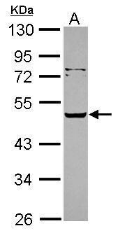
- Western Blot using SERPINA12 Polyclonal Antibody (Product # PA5-30989). Sample (30 µg of whole cell lysate). Lane A: Jurkat. 10% SDS PAGE. SERPINA12 Polyclonal Antibody (Product # PA5-30989) diluted at 1:5,000.
Supportive validation
- Submitted by
- Invitrogen Antibodies (provider)
- Main image

- Experimental details
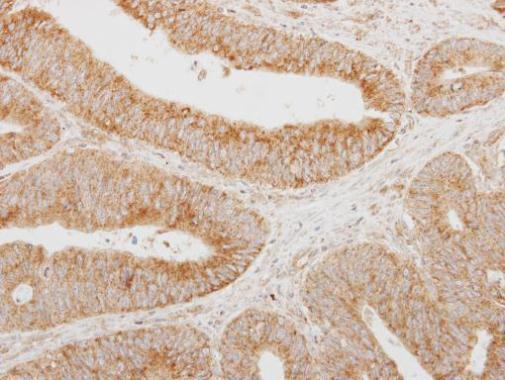
- Immunohistochemical analysis of paraffin-embedded human colon carcinoma, using SERPINA12 (Product # PA5-30989) antibody at 1:250 dilution. Antigen Retrieval: EDTA based buffer, pH 8.0, 15 min.
Supportive validation
- Submitted by
- Invitrogen Antibodies (provider)
- Main image
- Experimental details
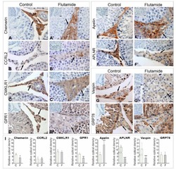
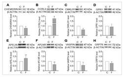
- Figure 1 Qualitative ( A - H , A' - H' ) and quantitative ( I ) analyses of immunohistochemical staining of chemerin ( A , A' ), CCRL2 ( B , B' ), CMKLR1 ( C , C' ), GPR1 ( D , D' ), apelin ( E , E' ), APLNR ( F , F' ), vaspin (G , G') and GRP78 (H , H') . Representative microphotographs of control (A - H) and flutamide-treated testes ( A' - H' ). Additionally stained with Mayer's haematoxylin. Bars = 10 mum. Positive staining for chemerin, CCRL2, CMKLR1, apelin and APLNR are restricted to Leydig cells (arrows), whereas staining of GPR1, vaspin and GRP78 is visible in Leydig cells (arrows) and seminiferous tubules (open arrows). Note the decreased levels of chemerin ( A' ), GPR1 ( D' ), APLNR ( F' ) and vaspin ( G' ) and increased levels of CCRL2 ( B' ) and apelin ( E' ) intensities after flutamide administration. The intensity of CMKLR1 and GRP78 staining in flutamide ( C',H' ) and control ( C , H ) Leydig cells are similar. No immunopositive staining of chemerin, apelin and vaspin and their receptors was observed when the primary antibodies are omitted ( A - H , A' - H', insertion in upper left corner of microphotographs). Histograms ( I ) depicting staining intensities of chemerin, apelin, vaspin, and their receptors are expressed as relative optical density of the brown staining product in Leydig cells. The results are presented as means +- SD. Statistically significant differences are flagged with asterisks (* p < 0.05, ** p < 0.01, *** p < 0.001). Control ( n = 6) and f

- Submitted by
- Invitrogen Antibodies (provider)
- Main image
- Experimental details
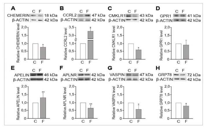
- Figure 2 Representative Western blots assessing the relative expression of chemerin ( A ), CCRL2 ( B ), CMKLR1 ( C ), GPR1 ( D ) , apelin ( E ), APLNR (F), vaspin ( G ) and GRP78 ( H ) proteins in control and flutamide-treated testes. Normalisation was performed with beta-Actin as a loading control. Protein levels within control testes were given a value of 1. Data obtained from three separate analyses are presented as means +- SD. Statistically significant differences are flagged with asterisks (* p < 0.05, ** p < 0.01) between control ( n = 6) and flutamide-treated ( n = 6) animals; C: control, F: flutamide.
- Submitted by
- Invitrogen Antibodies (provider)
- Main image

- Experimental details
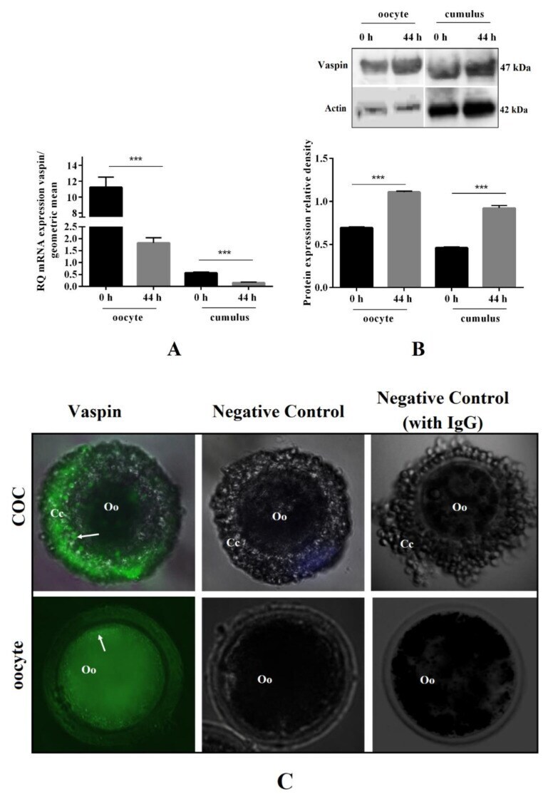
- Figure 1 Vaspin mRNA and protein expression before and after oocyte in vitro maturation, as well as its immunolocalisation in cumulus-oocyte complexes (COCs). COCs (50/group/experiment) were selected after morphological examination of material collected from the ovaries taken from 100 pigs (15 follicles/ovary). COCs were collected before (0 h) and after (44 h) in vitro maturation and oocytes were denuded by gently pipetting in hyaluronidase, then real-time PCR and Western blot analysis were performed to determine mRNA ( A ) and protein ( B ) level of vaspin in oocytes and cumulus cells. Additionally, immunolocalisation of vaspin was analysed by immunofluorescence in COCs ( C ). Gene expression level was normalised to the geometric mean of actin and glyceraldehyde 3-phosphate dehydrogenase (GAPDH), and protein to actin. Vaspin immunostaining shown by arrows. Experiments were performed independently and repeated three times ( n = 3 ). The data are plotted as the mean +- standard error of the mean (SEM) of three independent experiments. Significance between groups before and after maturation is indicated by *** p < 0.001; Cumulus cells (Cc), oocyte (Oo), Immunoglobulin G (IgG).