Explore
 Validate
 Learn
Learn Western blot
 Immunocytochemistry
Antibody data
- Antibody Data
- Antigen structure
- References [2]
- Comments [0]
- Validations
- Immunocytochemistry [1]
- Other assay [1]
Submit
Validation data
Reference
Comment
Report error
- Product number
- PA5-30869 - Provider product page

- Provider
- Invitrogen Antibodies
- Product name
- SGOL1 Polyclonal Antibody
- Antibody type
- Polyclonal
- Antigen
- Recombinant full-length protein
- Description
- Recommended positive controls: HeLa, HepG2. Store product as a concentrated solution. Centrifuge briefly prior to opening the vial.
- Reactivity
- Human
- Host
- Rabbit
- Isotype
- IgG
- Vial size
- 100 μL
- Concentration
- 1 mg/mL
- Storage
- Store at 4°C short term. For long term storage, store at -20°C, avoiding freeze/thaw cycles.
Submitted references Ginsenoside Rg1 suppresses cancer cell proliferation through perturbing mitotic progression.
Sensors at centrosomes reveal determinants of local separase activity.
Hong J, Gwon D, Jang CY
Journal of ginseng research 2022 May;46(3):481-488
Journal of ginseng research 2022 May;46(3):481-488
Sensors at centrosomes reveal determinants of local separase activity.
Agircan FG, Schiebel E
PLoS genetics 2014 Oct;10(10):e1004672
PLoS genetics 2014 Oct;10(10):e1004672
No comments: Submit comment
Supportive validation
- Submitted by
- Invitrogen Antibodies (provider)
- Main image
- Experimental details
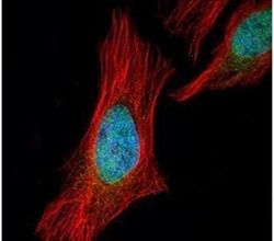
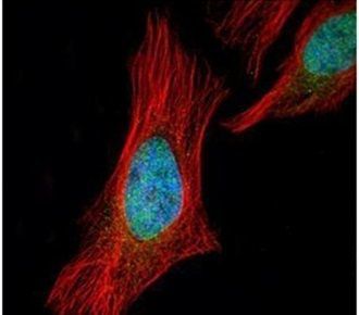
- Immunofluorescent analysis of SGOL1 in paraformaldehyde-fixed HeLa cells using a SGOL1 polyclonal antibody (Product # PA5-30869) (Green) at a 1:500 dilution. Alpha-tubulin filaments were labeled with Product # PA5-29281 (Red) at a 1:2000.
Supportive validation
- Submitted by
- Invitrogen Antibodies (provider)
- Main image
- Experimental details
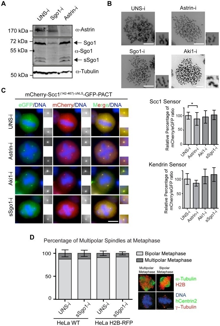
- Figure 3 Astrin (Spag5), Aki1 and Sgo1 depletion does not promote centriole disengagement. (A) Immunoblot analysis of HeLa cells following astrin and sSgo1/Sgo1 depletion. alpha-Tubulin was used as loading control. (B) Depletions of Sgo1, astrin or Aki1 caused premature loss of sister chromatid cohesin. Chromosome spreads of HeLa cells are shown. Four fold enlargements on the right show the separated sister chromatids. Scale bar: 10 um. (C) Representative images of HeLa cells stably expressing the Scc1 and kendrin sensors upon depletion of Sgo1, astrin or Aki1. The two-fold enlargements show the centrosomes. The relative mCherry/eGFP ratios upon siRNA treatment of cells are depicted on the right for the Scc1 (n>45) and kendrin (n>15) sensors. One-way ANOVA was used as statistical test (* represents p