Explore
 Validate
 Learn
Learn Western blot
 ELISA
ELISA Immunocytochemistry
 Immunohistochemistry
Antibody data
- Antibody Data
- Antigen structure
- References [0]
- Comments [0]
- Validations
- Western blot [1]
- Immunocytochemistry [6]
Submit
Validation data
Reference
Comment
Report error
- Product number
- LS-B1658 - Provider product page

- Provider
- LSBio
- Product name
- IHC-plus™ KRT18 / CK18 / Cytokeratin 18 Antibody (clone DA-7) LS-B1658
- Antibody type
- Monoclonal
- Description
- Affinity purified
- Reactivity
- Human
- Host
- Mouse
- Isotype
- IgG
- Antibody clone number
- DA-7
- Storage
- Store at 4°C. Avoid freezing.
No comments: Submit comment
Enhanced validation
- Submitted by
- LSBio (provider)
- Enhanced method
- Genetic validation
- Main image

- Experimental details
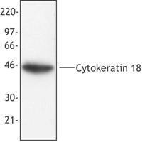
- MCF-7 cell extract was resolved by electrophoresis, transferred to nitrocellulose and probed with monoclonal anti-cytokeratin 18 antibody (clone DA-7). Proteins were visualized using a goat anti-mouse secondary conjugated to HRP and a chemiluminescence detection system.
Supportive validation
- Submitted by
- LSBio (provider)
- Enhanced method
- Genetic validation
- Main image

- Experimental details
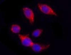
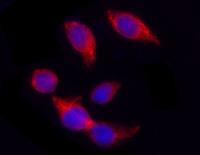
- Hela cells stained with purified mouse monoclonal antibody against Cytokeratin 18 (clone DA-7), followed by Rhodamine Red-X conjugated Donkey anti-mouse IgG and DAPI

- Submitted by
- LSBio (provider)
- Enhanced method
- Genetic validation
- Main image

- Experimental details
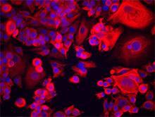
- MCF-7 cells were stained with anti-Cytokeratin 18 (clone DA-7), followed by Alexa Fluor 546 secondary antibody and DAPI (nuclei). Images were acquired on a Nikon FC300 inverted microscope at 20X magnification. Data provided by Dr. John Nolan, La Jolla Bioengineering Institute.

- Submitted by
- LSBio (provider)
- Main image
- Experimental details
- Hela cells stained with purified mouse monoclonal antibody against Cytokeratin 18 (clone DA-7), followed by Rhodamine Red-X conjugated Donkey anti-mouse IgG and DAPI

- Submitted by
- LSBio (provider)
- Main image

- Experimental details
- MCF-7 cells were stained with anti-Cytokeratin 18 (clone DA-7), followed by Alexa Fluor 546 secondary antibody and DAPI (nuclei). Images were acquired on a Nikon FC300 inverted microscope at 20X magnification. Data provided by Dr. John Nolan, La Jolla Bioengineering Institute.

- Submitted by
- LSBio (provider)
- Main image

- Experimental details
- Hela cells stained with purified mouse monoclonal antibody against Cytokeratin 18 (clone DA-7), followed by Rhodamine Red-X conjugated Donkey anti-mouse IgG and DAPI
- Submitted by
- LSBio (provider)
- Main image

- Experimental details
- MCF-7 cells were stained with anti-Cytokeratin 18 (clone DA-7), followed by Alexa Fluor 546 secondary antibody and DAPI (nuclei). Images were acquired on a Nikon FC300 inverted microscope at 20X magnification. Data provided by Dr. John Nolan, La Jolla Bioengineering Institute.