Explore
 Validate
 Learn
Learn Western blot
 ELISA
ELISAAntibody data
- Antibody Data
- Antigen structure
- References [0]
- Comments [0]
- Validations
- Western blot [3]
- Immunohistochemistry [13]
Submit
Validation data
Reference
Comment
Report error
- Product number
- STJ92631 - Provider product page

- Provider
- St John's Laboratory
- Product name
- Anti-KRT18 antibody (10-90) (STJ92631)
- Antibody type
- Polyclonal
- Description
- Rabbit polyclonal antibody anti-Keratin-Type I Cytoskeletal 18 (10-90) is suitable for use in Western Blot, Immunohistochemistry, Immunoprecipitation, Immunofluorescence, Immunocytochemistry and ELISA research applications.
- Reactivity
- Human, Mouse, Rat
- Host
- Rabbit
- Conjugate
- Unconjugated
- Antigen sequence
NA- Epitope
- NA
- Isotype
- IgG
- Antibody clone number
- NA
- Vial size
- NA
- Concentration
- NA
- Storage
- Store at-20°C for up to 1 year from the date of receipt, and avoid repeat freeze-thaw cycles.
- Handling
- NA
No comments: Submit comment
Supportive validation
Supportive validation
Supportive validation
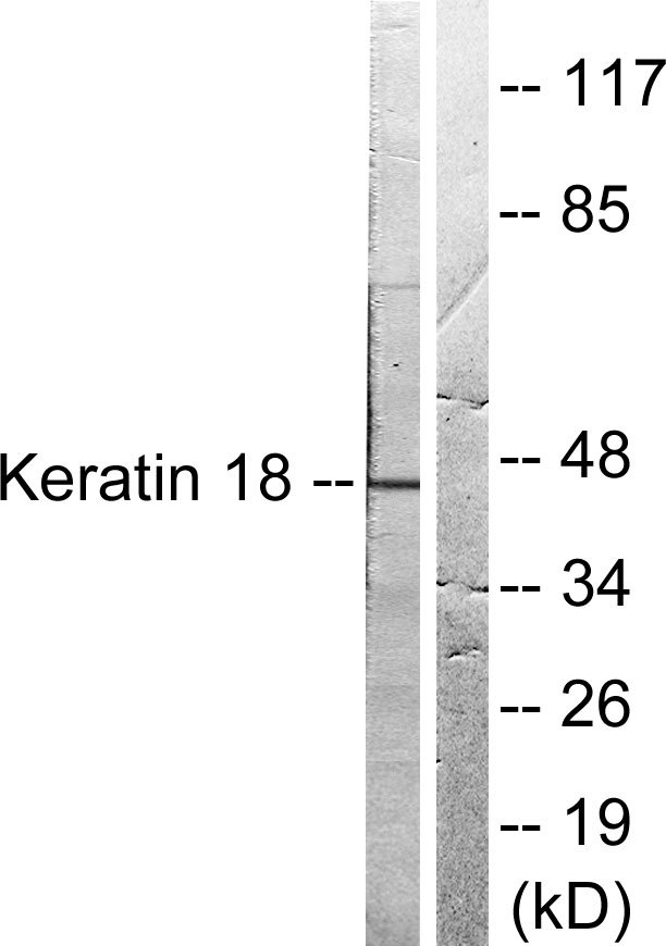
- Submitted by
- St John's Laboratory (provider)
- Main image

- Experimental details
- Western blot analysis of lysates from HeLa cells, treated with UV 5', using Keratin 18 Antibody. The lane on the right is blocked with the synthesized peptide.
- Sample type
- NA
- Validation comment
- NA
- Primary Ab dilution
- NA
- Other comments
- NA
- Secondary Ab
- NA
- Secondary Ab dilution
- NA
- Protocol
- NA
Supportive validation
- Submitted by
- St John's Laboratory (provider)
- Main image
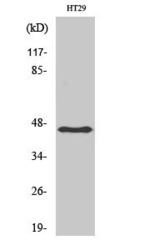
- Experimental details
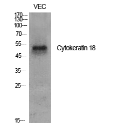
- Western blot analysis of HT29 cells using Cytokeratin 18 Polyclonal Antibody diluted at 1ï¼2000
- Sample type
- NA
- Validation comment
- NA
- Primary Ab dilution
- NA
- Other comments
- NA
- Secondary Ab
- NA
- Secondary Ab dilution
- NA
- Protocol
- NA
Supportive validation
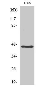
- Submitted by
- St John's Laboratory (provider)
- Main image

- Experimental details
- Western blot analysis of various cells using Cytokeratin 18 Polyclonal Antibody diluted at 1ï¼2000
- Sample type
- NA
- Validation comment
- NA
- Primary Ab dilution
- NA
- Other comments
- NA
- Secondary Ab
- NA
- Secondary Ab dilution
- NA
- Protocol
- NA
Supportive validation
Supportive validation
Supportive validation
Supportive validation
Supportive validation
Supportive validation
Supportive validation
Supportive validation
Supportive validation
Supportive validation
Supportive validation
Supportive validation
Supportive validation
- Submitted by
- St John's Laboratory (provider)
- Main image

- Experimental details
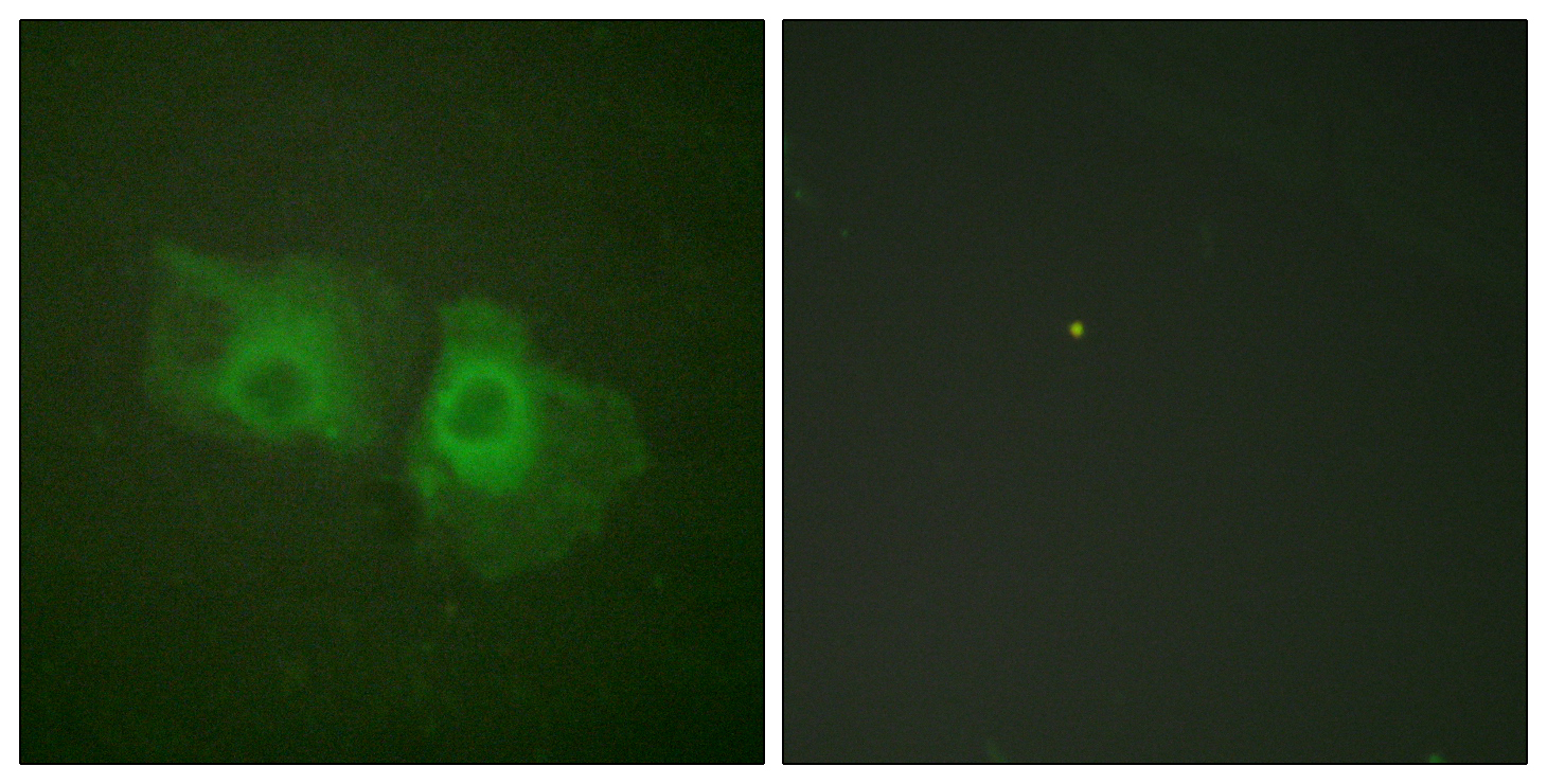
- Immunofluorescence analysis of HeLa cells, using Keratin 18 Antibody. The picture on the right is blocked with the synthesized peptide.
- Sample type
- NA
- Validation comment
- NA
- Primary Ab dilution
- NA
- Other comments
- NA
- Secondary Ab
- NA
- Secondary Ab dilution
- NA
- Protocol
- NA
Supportive validation
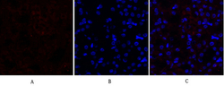
- Submitted by
- St John's Laboratory (provider)
- Main image
- Experimental details
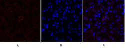
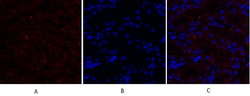
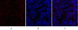
- Immunofluorescence analysis of mouse-kidney tissue. 1, Cytokeratin 18 Polyclonal Antibody (red) was diluted at 1:200 (4°C, overnight). 2, Cy3 labled Secondary antibody was diluted at 1:300 (room temperature, 50min).3, Picture B: DAPI (blue) 10min. Picture A:Target. Picture B: DAPI. Picture C: merge of A+B
- Sample type
- NA
- Validation comment
- NA
- Primary Ab dilution
- NA
- Other comments
- NA
- Secondary Ab
- NA
- Secondary Ab dilution
- NA
- Protocol
- NA
Supportive validation
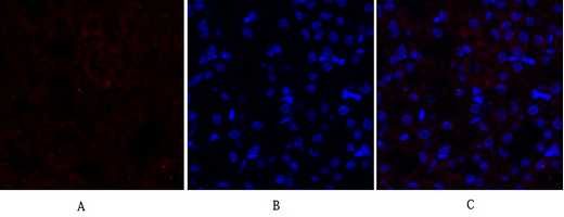
- Submitted by
- St John's Laboratory (provider)
- Main image
- Experimental details
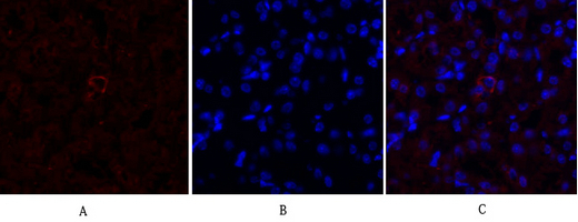
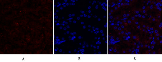
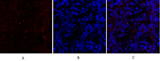
- Immunofluorescence analysis of mouse-kidney tissue. 1, Cytokeratin 18 Polyclonal Antibody (red) was diluted at 1:200 (4°C, overnight). 2, Cy3 labled Secondary antibody was diluted at 1:300 (room temperature, 50min).3, Picture B: DAPI (blue) 10min. Picture A:Target. Picture B: DAPI. Picture C: merge of A+B
- Sample type
- NA
- Validation comment
- NA
- Primary Ab dilution
- NA
- Other comments
- NA
- Secondary Ab
- NA
- Secondary Ab dilution
- NA
- Protocol
- NA
Supportive validation
- Submitted by
- St John's Laboratory (provider)
- Main image
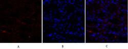
- Experimental details
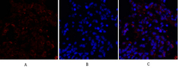
- Immunofluorescence analysis of rat-kidney tissue. 1, Cytokeratin 18 Polyclonal Antibody (red) was diluted at 1:200 (4°C, overnight). 2, Cy3 labled Secondary antibody was diluted at 1:300 (room temperature, 50min).3, Picture B: DAPI (blue) 10min. Picture A:Target. Picture B: DAPI. Picture C: merge of A+B
- Sample type
- NA
- Validation comment
- NA
- Primary Ab dilution
- NA
- Other comments
- NA
- Secondary Ab
- NA
- Secondary Ab dilution
- NA
- Protocol
- NA
Supportive validation
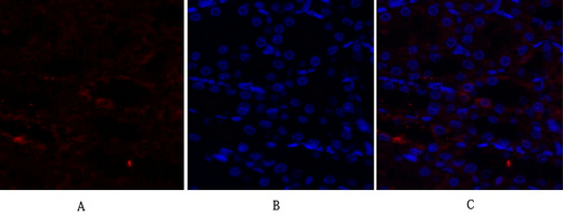
- Submitted by
- St John's Laboratory (provider)
- Main image
- Experimental details
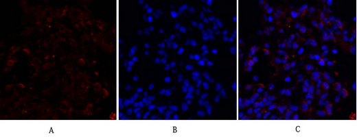
- Immunofluorescence analysis of rat-kidney tissue. 1, Cytokeratin 18 Polyclonal Antibody (red) was diluted at 1:200 (4°C, overnight). 2, Cy3 labled Secondary antibody was diluted at 1:300 (room temperature, 50min).3, Picture B: DAPI (blue) 10min. Picture A:Target. Picture B: DAPI. Picture C: merge of A+B
- Sample type
- NA
- Validation comment
- NA
- Primary Ab dilution
- NA
- Other comments
- NA
- Secondary Ab
- NA
- Secondary Ab dilution
- NA
- Protocol
- NA
Supportive validation
- Submitted by
- St John's Laboratory (provider)
- Main image
- Experimental details
- Immunofluorescence analysis of rat-lung tissue. 1, Cytokeratin 18 Polyclonal Antibody (red) was diluted at 1:200 (4°C, overnight). 2, Cy3 labled Secondary antibody was diluted at 1:300 (room temperature, 50min).3, Picture B: DAPI (blue) 10min. Picture A:Target. Picture B: DAPI. Picture C: merge of A+B
- Sample type
- NA
- Validation comment
- NA
- Primary Ab dilution
- NA
- Other comments
- NA
- Secondary Ab
- NA
- Secondary Ab dilution
- NA
- Protocol
- NA
Supportive validation
- Submitted by
- St John's Laboratory (provider)
- Main image
- Experimental details
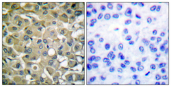
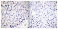
- Immunohistochemistry analysis of paraffin-embedded human breast carcinoma tissue, using Keratin 18 Antibody. The picture on the right is blocked with the synthesized peptide.
- Sample type
- NA
- Validation comment
- NA
- Primary Ab dilution
- NA
- Other comments
- NA
- Secondary Ab
- NA
- Secondary Ab dilution
- NA
- Protocol
- NA
Supportive validation
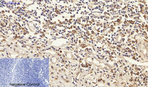
- Submitted by
- St John's Laboratory (provider)
- Main image
- Experimental details
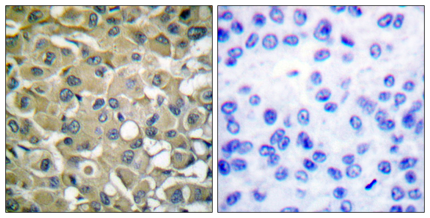
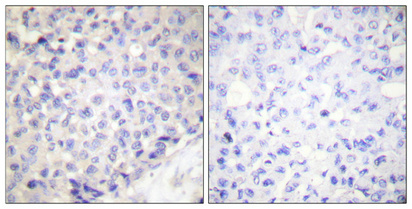
- Immunohistochemical analysis of paraffin-embedded Human breast cancer. Antibody was diluted at 1:100 (4°C overnight). High-pressure and temperature Tris-EDTA, pH8.0 was used for antigen retrieval. Negetive contrl (right) obtaned from antibody was pre-absorbed by immunogen peptide.
- Sample type
- NA
- Validation comment
- NA
- Primary Ab dilution
- NA
- Other comments
- NA
- Secondary Ab
- NA
- Secondary Ab dilution
- NA
- Protocol
- NA
Supportive validation
- Submitted by
- St John's Laboratory (provider)
- Main image
- Experimental details
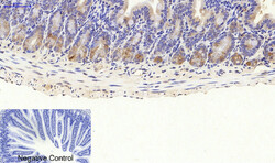
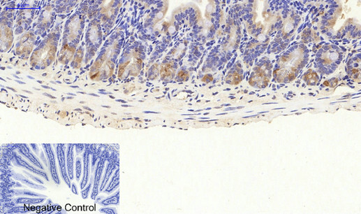
- Immunohistochemical analysis of paraffin-embedded Mouse-colon tissue. 1, CytokeRatin 18 Polyclonal Antibody was diluted at 1:200 (4°C, overnight). 2, Sodium citrate pH 6.0 was used for antibody retrieval (>98°C, 20min). 3, Secondary antibody was diluted at 1:200 (room tempeRature, 30min). Negative control was used by secondary antibody only.
- Sample type
- NA
- Validation comment
- NA
- Primary Ab dilution
- NA
- Other comments
- NA
- Secondary Ab
- NA
- Secondary Ab dilution
- NA
- Protocol
- NA
Supportive validation
- Submitted by
- St John's Laboratory (provider)
- Main image
- Experimental details
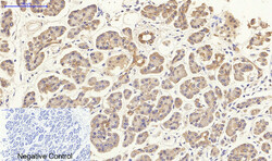
- Immunohistochemical analysis of paraffin-embedded Human-stomach-cancer tissue. 1, CytokeRatin 18 Polyclonal Antibody was diluted at 1:200 (4°C, overnight). 2, Sodium citrate pH 6.0 was used for antibody retrieval (>98°C, 20min). 3, Secondary antibody was diluted at 1:200 (room tempeRature, 30min). Negative control was used by secondary antibody only.
- Sample type
- NA
- Validation comment
- NA
- Primary Ab dilution
- NA
- Other comments
- NA
- Secondary Ab
- NA
- Secondary Ab dilution
- NA
- Protocol
- NA

Supportive validation
- Submitted by
- St John's Laboratory (provider)
- Main image
- Experimental details
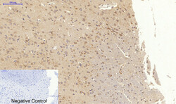
- Immunohistochemical analysis of paraffin-embedded Rat-brain tissue. 1, CytokeRatin 18 Polyclonal Antibody was diluted at 1:200 (4°C, overnight). 2, Sodium citrate pH 6.0 was used for antibody retrieval (>98°C, 20min). 3, Secondary antibody was diluted at 1:200 (room tempeRature, 30min). Negative control was used by secondary antibody only.
- Sample type
- NA
- Validation comment
- NA
- Primary Ab dilution
- NA
- Other comments
- NA
- Secondary Ab
- NA
- Secondary Ab dilution
- NA
- Protocol
- NA
Supportive validation
- Submitted by
- St John's Laboratory (provider)
- Main image

- Experimental details
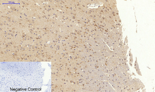
- Immunohistochemical analysis of paraffin-embedded Human-Tonsil tissue. 1, CytokeRatin 18 Polyclonal Antibody was diluted at 1:200 (4°C, overnight). 2, Sodium citrate pH 6.0 was used for antibody retrieval (>98°C, 20min). 3, Secondary antibody was diluted at 1:200 (room tempeRature, 30min). Negative control was used by secondary antibody only.
- Sample type
- NA
- Validation comment
- NA
- Primary Ab dilution
- NA
- Other comments
- NA
- Secondary Ab
- NA
- Secondary Ab dilution
- NA
- Protocol
- NA