Explore
 Validate
 Learn
Learn Western blot
 ELISA
ELISAAntibody data
- Antibody Data
- Antigen structure
- References [0]
- Comments [0]
- Validations
- Western blot [2]
- Immunocytochemistry [2]
- Immunohistochemistry [3]
- Flow cytometry [2]
Submit
Validation data
Reference
Comment
Report error
- Product number
- F50352 - Provider product page

- Provider
- NSJ Bioreagents
- Product name
- Cytokeratin-18 Antibody
- Antibody type
- Polyclonal
- Description
- This highly specific Cytokeratin-18 antibody is suitable for use in Flow cytometry/Immunofluorescence/Immunohistochemistry/Western blot/ELISA applications with human and mouse samples.
- Reactivity
- Human, Mouse
- Host
- Rabbit
- Conjugate
- Unconjugated
- Vial size
- 0.08 ml, 0.4 ml
- Concentration
- In 1X PBS, pH 7.4, with 0.09% sodium azide
- Storage
- Aliquot the Cytokeratin-18 antibody and store frozen at -20oC or colder. Avoid repeated freeze-thaw cycles.
No comments: Submit comment
Supportive validation
- Submitted by
- NSJ Bioreagents (provider)
- Main image
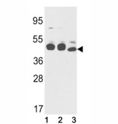
- Experimental details
- Western blot analysis of Cytokeratin-18 antibody and (1) K562, (2) NCI-H460, and (3) mouse stomach lysate.

- Submitted by
- NSJ Bioreagents (provider)
- Main image

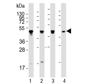
- Experimental details
- Western blot testing of human 1) NCI-H460, 2) K562, 3) Hela and 4) A431 whole cell lysate with Cytokeratin-18 antibody at 1:2000. Predicted molecular weight ~48 kDa.
Supportive validation
- Submitted by
- NSJ Bioreagents (provider)
- Main image
- Experimental details
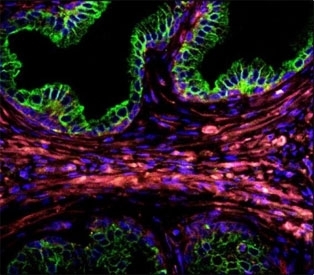
- Confocal immunofluorescent analysis of Cytokeratin-18 antibody with prostate carcinoma followed by Alexa Fluor 488-conjugated goat anti-rabbit lgG (green). DAPI was used as a nuclear counterstain (blue).
- Submitted by
- NSJ Bioreagents (provider)
- Main image
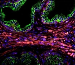
- Experimental details
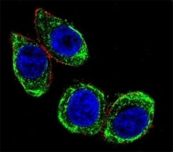
- Confocal immunofluorescent analysis of Cytokeratin-18 antibody with HeLa cells followed by Alexa Fluor 488-conjugated goat anti-rabbit lgG (green). Actin filaments have been labeled with Alexa Fluor 555 Phalloidin (red). DAPI was used as a nuclear counterstain (blue).

Supportive validation
- Submitted by
- NSJ Bioreagents (provider)
- Main image

- Experimental details
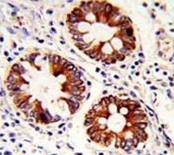
- IHC analysis of FFPE human colon carcinoma with Cytokeratin-18 antibody
- Submitted by
- NSJ Bioreagents (provider)
- Main image
- Experimental details
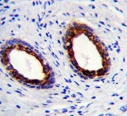
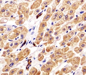
- Cytokeratin-18 antibody immunohistochemistry analysis in formalin fixed and paraffin embedded human prostate carcinoma.

- Submitted by
- NSJ Bioreagents (provider)
- Main image
- Experimental details
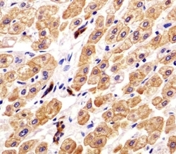
- IHC analysis of FFPE human liver tissue with Cytokeratin-18 antibody. Required HIER: steamed antigen retrieval with pH6 citrate buffer.
Supportive validation
- Submitted by
- NSJ Bioreagents (provider)
- Main image
- Experimental details
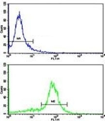
- Flow cytometric analysis of WiDr cells using Cytokeratin-18 antibody (bottom histogram) compared to a negative control (top histogram). FITC-conjugated goat-anti-rabbit secondary Ab was used for the analysis.
- Submitted by
- NSJ Bioreagents (provider)
- Main image
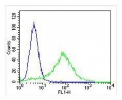
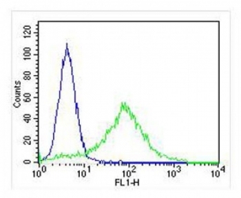
- Experimental details
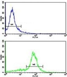
- FACS testing of fixed and permeabilized human HeLa cells with Cytokeratin-18 antibody (green) at 1:25 and negative control (blue).