 Explore
 Validate
 Learn
Learn Western blot
 Immunocytochemistry
Antibody data
- Antibody Data
- Antigen structure
- References [0]
- Comments [0]
- Validations
- Western blot [2]
- Immunohistochemistry [1]
Submit
Validation data
Reference
Comment
Report error
- Product number
- NBP2-34356-0.1mg - Provider product page

- Provider
- Novus Biologicals
- Product name
- Mouse Monoclonal Cytokeratin 18 Antibody
- Antibody type
- Monoclonal
- Description
- Protein G purified. This MAb reacts with a wide variety of simple epithelia. It does not react with stratified squamous epithelia. It reacts with epithelial tumors of the gastrointestinal tract, lung, breast, pancreas, ovary, and thyroid. Cytokeratin 18, which belongs to the type A (acidic) subfamily of low molecular weight keratins, exists in combination with cytokeratin 8. It is reported that tissues from gastrointestinal tract are positive for both cytokeratin 8 and 18 but do not contain cytokeratin 14. Tissues from gastrointestinal tract, respiratory tract and urogenital tract, as well as endocrine and exocrine tissues and mesothelial cells are positive for cytokeratin 18.
- Reactivity
- Human, Mouse, Rat, Bovine, Canine, Hamster, Porcine, Sheep
- Host
- Mouse
- Isotype
- IgG
- Vial size
- 0.1 mg
- Concentration
- 0.2 mg/ml
- Storage
- Store at 4C.
No comments: Submit comment
Supportive validation
- Submitted by
- Novus Biologicals (provider)
- Main image
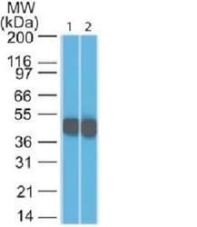
- Experimental details
- Western Blot: Cytokeratin 18 Antibody (SPM265) [NBP2-34356] - Western Blot 1) HeLa and 2) A431 Lysate using Cytokeratin 18 Monoclonal Antibody (SPM265)

- Submitted by
- Novus Biologicals (provider)
- Main image
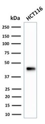
- Experimental details
- Western Blot: Cytokeratin 18 Antibody (SPM265) [NBP2-34356] - Western Blot Analysis of HCT116 cell lysate using Cytokeratin 18 Antibody (SPM265).

Supportive validation
- Submitted by
- Novus Biologicals (provider)
- Main image
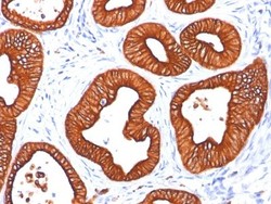
- Experimental details
- Immunohistochemistry-Paraffin: Cytokeratin 18 Antibody (SPM265) [NBP2-34356] - Formalin-paraffin skin sweat gland stained with Cytokeratin 18 MAb (SPM265). Note cytoplasmic staining of tumor cells.
