Explore
 Validate
 Learn
Learn Western blot
 Immunocytochemistry
 Immunohistochemistry
Antibody data
- Antibody Data
- Antigen structure
- References [0]
- Comments [0]
- Validations
- Immunocytochemistry [3]
Submit
Validation data
Reference
Comment
Report error
- Product number
- 14-9815-37 - Provider product page

- Provider
- Invitrogen Antibodies
- Product name
- Cytokeratin 18 Monoclonal Antibody (LDK18), eBioscience™
- Antibody type
- Monoclonal
- Antigen
- Other
- Description
- Description: The monoclonal antibody LDK18 recognizes human Cytokeratin 18. Cytokeratin 18 is member of the large family of intermediate filaments found within epithelial cells. The family of cytokeratins is made up of type I (acidic) and type II (basic) members which form heterodimers by pairing an acidic with a basic member to compose the keratin filament. Cytokeratin 18 (type I) is most commonly found associated with cytokeratin 8 (type II). Because many tumors of epithelial origin retain expression of keratin proteins, cytokeratin 18 has been used as a tumor marker to identify both tumors and metastases. In addition, cytokeratin 18 has been shown to be expressed in highly proliferative retinal pigment epithelium cells. Applications Reported: This LDK18 antibody has been reported for use in western blotting, microscopy, immunohistochemical staining, and immunocytochemistry. Applications Tested: This LDK18 antibody has been tested by immmunocytochemistry of MeOH-fixed human cells and can be used at less than or equal to 1 µg/mL. It is recommended that the antibody be titrated for optimal performance in the assay of interest. Purity: Greater than 90%, as determined by SDS-PAGE. Aggregation: Less than 10%, as determined by HPLC. Filtration: 0.2 µm post-manufacturing filtered.
- Reactivity
- Human
- Host
- Mouse
- Isotype
- IgG
- Antibody clone number
- LDK18
- Vial size
- 2 mg
- Concentration
- 0.5 mg/mL
- Storage
- 4°C
No comments: Submit comment
Supportive validation
- Submitted by
- Invitrogen Antibodies (provider)
- Main image

- Experimental details
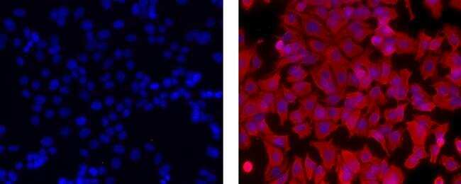
- Immunocytochemistry of fixed MCF-7 cells stained with 1 µg/mL Mouse IgG1 K Isotype Control Purified (left) or 1 µg/mL Anti-Human Cytokeratin 18 Purified (right) followed by F (ab')2 Anti-Mouse IgG eFluor® 570 (Product # 41-4010-82).Nuclei are stained with DAPI.
- Submitted by
- Invitrogen Antibodies (provider)
- Main image
- Experimental details
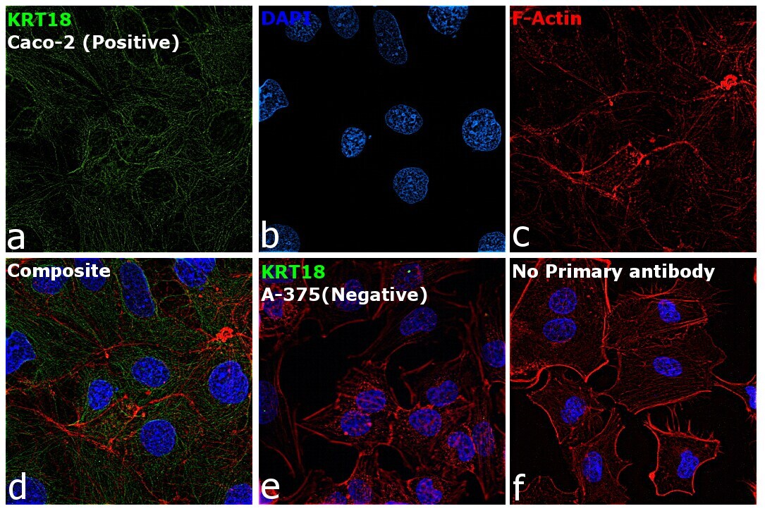
- Immunofluorescence analysis of Cytokeratin 18 was performed using 70% confluent log phase Caco-2 cells. The cells were fixed with ice-cold acetone at 4°C for 5 minutes and blocked with 2% BSA for 45 minutes at room temperature. The cells were labeled with Cytokeratin 18 Monoclonal Antibody (LDK18), eBioscience™ (Product # 14-9815-82) at 5 µg/mL in 0.1% BSA, incubated at 4 degree celsius overnight and then labeled with Goat anti-Mouse IgG (H+L) Superclonal™ Recombinant Secondary Antibody, Alexa Fluor® 488 conjugate (Product # A28175), (1:2000 dilution), for 45 minutes at room temperature (Panel a: Green). Nuclei (Panel b: Blue) were stained with SlowFade® Gold Antifade Mountant with DAPI (Product # S36938). F-actin (Panel c: Red) was stained with Rhodamine Phalloidin (Product # R415, 1:300 dilution). Panel d represents the merged image showing cytoskeletal localization. Panel e represents A-375. Panel f represents control cells with no primary antibody to assess background. The images were captured at 60X magnification.

- Submitted by
- Invitrogen Antibodies (provider)
- Main image

- Experimental details
- Immunofluorescence analysis of Cytokeratin 18 was performed using 70% confluent log phase Caco-2 cells. The cells were fixed with ice-cold acetone at 4°C for 5 minutes and blocked with 2% BSA for 45 minutes at room temperature. The cells were labeled with Cytokeratin 18 Monoclonal Antibody (LDK18), eBioscience™ (Product # 14-9815-82) at 5 µg/mL in 0.1% BSA, incubated at 4 degree celsius overnight and then labeled with Goat anti-Mouse IgG (H+L) Superclonal™ Recombinant Secondary Antibody, Alexa Fluor® 488 conjugate (Product # A28175), (1:2000 dilution), for 45 minutes at room temperature (Panel a: Green). Nuclei (Panel b: Blue) were stained with SlowFade® Gold Antifade Mountant with DAPI (Product # S36938). F-actin (Panel c: Red) was stained with Rhodamine Phalloidin (Product # R415, 1:300 dilution). Panel d represents the merged image showing cytoskeletal localization. Panel e represents A-375. Panel f represents control cells with no primary antibody to assess background. The images were captured at 60X magnification.