Explore
 Validate
 Learn
Learn Western blot
 Immunocytochemistry
Antibody data
- Antibody Data
- Antigen structure
- References [5]
- Comments [0]
- Validations
- Western blot [1]
- Immunohistochemistry [1]
- Flow cytometry [1]
- Other assay [1]
Submit
Validation data
Reference
Comment
Report error
- Product number
- MA5-12281 - Provider product page

- Provider
- Invitrogen Antibodies
- Product name
- Anti-Cytokeratin 8/18 Antibody Cocktail
- Antibody type
- Monoclonal
- Antigen
- Purifed from natural sources
- Description
- MA5-12281 targets Cytokeratin 8/18 in IHC (P) applications and shows reactivity with Human samples. The MA5-12281 immunogen is semi-purified human cytokeratin preparation.
- Reactivity
- Human
- Host
- Mouse
- Isotype
- IgG
- Antibody clone number
- K8.8 + DC10
- Vial size
- 500 µL
- Concentration
- 0.2 mg/mL
- Storage
- 4° C
Submitted references The Hippo kinases LATS1 and 2 control human breast cell fate via crosstalk with ERα.
Relationship of CK8/18 expression pattern to breast cancer immunohistochemical subtyping in Egyptian patients.
Expression of KRT7 and WT1 differentiates precursor lesions of Wilms' tumours from those of papillary renal cell tumours and mucinous tubular and spindle cell carcinomas.
Compensation of type I and type II cytokeratin pools in lung cancer.
Patterns of nestin and other intermediate filament expression distinguish between gastrointestinal stromal tumors, leiomyomas and schwannomas.
Britschgi A, Duss S, Kim S, Couto JP, Brinkhaus H, Koren S, De Silva D, Mertz KD, Kaup D, Varga Z, Voshol H, Vissieres A, Leroy C, Roloff T, Stadler MB, Scheel CH, Miraglia LJ, Orth AP, Bonamy GM, Reddy VA, Bentires-Alj M
Nature 2017 Jan 26;541(7638):541-545
Nature 2017 Jan 26;541(7638):541-545
Relationship of CK8/18 expression pattern to breast cancer immunohistochemical subtyping in Egyptian patients.
Aiad HA, Samaka RM, Asaad NY, Kandil MA, Shehata MA, Miligy IM
Ecancermedicalscience 2014;8:404
Ecancermedicalscience 2014;8:404
Expression of KRT7 and WT1 differentiates precursor lesions of Wilms' tumours from those of papillary renal cell tumours and mucinous tubular and spindle cell carcinomas.
Szponar A, Kovacs G
Virchows Archiv : an international journal of pathology 2012 Apr;460(4):423-7
Virchows Archiv : an international journal of pathology 2012 Apr;460(4):423-7
Compensation of type I and type II cytokeratin pools in lung cancer.
Kanaji N, Bandoh S, Fujita J, Ishii T, Ishida T, Kubo A
Lung cancer (Amsterdam, Netherlands) 2007 Mar;55(3):295-302
Lung cancer (Amsterdam, Netherlands) 2007 Mar;55(3):295-302
Patterns of nestin and other intermediate filament expression distinguish between gastrointestinal stromal tumors, leiomyomas and schwannomas.
Sarlomo-Rikala M, Tsujimura T, Lendahl U, Miettinen M
APMIS : acta pathologica, microbiologica, et immunologica Scandinavica 2002 Jun;110(6):499-507
APMIS : acta pathologica, microbiologica, et immunologica Scandinavica 2002 Jun;110(6):499-507
No comments: Submit comment
Supportive validation
- Submitted by
- Invitrogen Antibodies (provider)
- Main image

- Experimental details
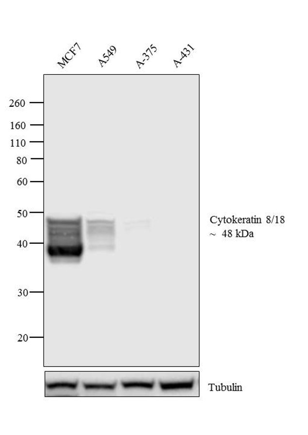
- Western blot analysis was performed on membrane enriched extracts (30 µg lysate) of MCF7 (Lane 1), A549 (Lane 2), A-375 (Lane 3) and A-431 (Lane 4). The blot was probed with Anti- Cytokeratin 8/18 Mouse Antibody Cocktail (Product # MA5-12281, 1 µg/mL) and detected by chemiluminescence using Goat anti-Mouse IgG (H+L) Superclonal™ Secondary Antibody, HRP conjugate (Product # A28177, 0.25 µg/mL, 1:4000 dilution). A 48 kDa band corresponding to Cytokeratin 8/18 was observed in MCF7, A549 and not observed in other cell lines which are documented to be Cytokeratin 8/18 negative.
Supportive validation
- Submitted by
- Invitrogen Antibodies (provider)
- Main image
- Experimental details
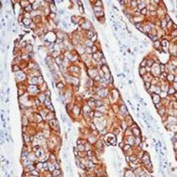
- Formalin-fixed, paraffin-embedded human breast cancer stained with Keratin, 8/18 antibody using peroxidase-conjugate and DAB chromogen. Note cytoplasmic staining of tumor cells.

Supportive validation
- Submitted by
- Invitrogen Antibodies (provider)
- Main image
- Experimental details
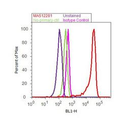
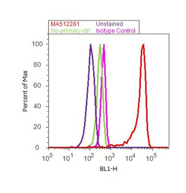
- Flow cytometry analysis of Cytokeratin 8, 18 was done on MCF7 cells. Cells were fixed with 70% ethanol for 10 minutes, permeabilized with 0.25% Triton™ X-100 for 20 minutes, and blocked with 5% BSA for 30 minutes at room temperature. Cells were labeled with Cytokeratin 8, 18 Mouse Monoclonal Antibody (MA5-12281, red histogram) or with mouse isotype control (pink histogram) at 3-5 ug/million cells in 2.5% BSA. After incubation at room temperature for 2 hours, the cells were labeled with Alexa Fluor® 488 Rabbit Anti-Mouse Secondary Antibody (A11059) at a dilution of 1:400 for 30 minutes at room temperature. The representative 10, 000 cells were acquired and analyzed for each sample using an Attune® Acoustic Focusing Cytometer. The purple histogram represents unstained control cells and the green histogram represents no-primary-antibody control.
Supportive validation
- Submitted by
- Invitrogen Antibodies (provider)
- Main image

- Experimental details
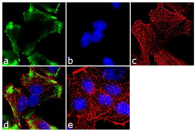
- Immunofluorescent analysis of Cytokeratin 8, 18 was performed using 70% confluent log phase MCF-7 cells. The cells were fixed with 4% paraformaldehyde for 10 minutes, permeabilized with 0.1% Triton™ X-100 for 10 minutes, and blocked with 1% BSA for 1 hour at room temperature. The cells were labeled with Cytokeratin 8, 18 (K8.8 + DC10) Mouse Monoclonal Antibody (Product # MA5-12281) at 2 µg/mL in 0.1% BSA and incubated for 3 hours at room temperature and then labeled with Goat anti-Mouse IgG (H+L) Superclonal™ Secondary Antibody, Alexa Fluor® 488 conjugate (Product # A28175) a dilution of 1:2000 for 45 minutes at room temperature (Panel a: green). Nuclei (Panel b: blue) were stained with SlowFade® Gold Antifade Mountant with DAPI (Product # S36938). F-actin (Panel c: red) was stained with Alexa Fluor® 555 Rhodamine Phalloidin (Product # R415, 1:300). Panel d represents the merged image showing cytoplasmic localization. Panel e shows the no primary antibody control. The images were captured at 60X magnification.