Explore
 Validate
 Learn
Learn Western blot
 ELISA
ELISAAntibody data
- Antibody Data
- Antigen structure
- References [0]
- Comments [0]
- Validations
- Western blot [3]
- Immunohistochemistry [1]
Submit
Validation data
Reference
Comment
Report error
- Product number
- MA5-15458 - Provider product page

- Provider
- Invitrogen Antibodies
- Product name
- Cytokeratin 18 Monoclonal Antibody (4D11E4)
- Antibody type
- Monoclonal
- Antigen
- Purifed from natural sources
- Description
- MA5-15458 targets Cytokeratin 18 in indirect ELISA, IHC and WB applications and shows reactivity with Human samples. The MA5-15458 immunogen is purified recombinant fragment of human Cytokeratin 18 (aa391-483) expressed in E. Coli.. MA5-15458 detects Cytokeratin 18 which has a predicted molecular weight of approximately 48kDa.
- Reactivity
- Human, Mouse
- Host
- Mouse
- Isotype
- IgG
- Antibody clone number
- 4D11E4
- Vial size
- 100 µL
- Concentration
- Conc. Not Determined
- Storage
- Store at 4°C short term. For long term storage, store at -20°C, avoiding freeze/thaw cycles.
No comments: Submit comment
Supportive validation
- Submitted by
- Invitrogen Antibodies (provider)
- Main image

- Experimental details
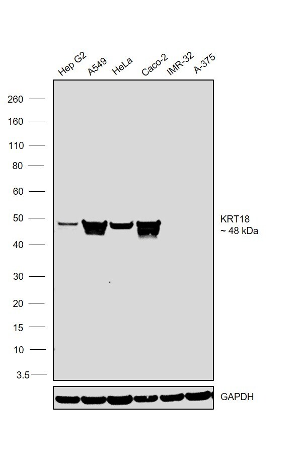
- Western blot analysis of Cytokeratin 18 using Cytokeratin 18 monoclonal antibody (Product # MA5-15458) in HeLa (1), NIH/3T3 (2), A549 (3), Jurkat (4), MCF-7 (5), HepG2 (6), A431 (7), HEK293 (8) and K562 (9) cell lysate.

- Submitted by
- Invitrogen Antibodies (provider)
- Main image

- Experimental details
- Western blot analysis of Cytokeratin 18 using Cytokeratin 18 monoclonal antibody (Product # MA5-15458) in HeLa (1), NIH/3T3 (2), A549 (3), Jurkat (4), MCF-7 (5), HepG2 (6), A431 (7), HEK293 (8) and K562 (9) cell lysate.
- Submitted by
- Invitrogen Antibodies (provider)
- Main image

- Experimental details
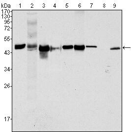
- Western blot was performed using Cytokeratin 18 Monoclonal Antibody (4D11E4) (Product # MA5-15458) and a 48kDa band corresponding to KRT18 was observed across all the cell lines tested expect IMR-32 and A-375. Whole cell extracts (30 µg lysate) of Hep G2 (Lane 1), A549 (Lane 2), HeLa (Lane 3), Caco-2 (Lane 4), IMR-32 (Lane 5) and A-375 (Lane 6) were electrophoresed using NuPAGE™ 4-12% Bis-Tris Protein Gel (Product # NP0322BOX). Resolved proteins were then transferred onto a nitrocellulose membrane (Product # IB23001) by iBlot® 2 Dry Blotting System (Product # IB21001). The blot was probed with the primary antibody (1:1000 dilution) and detected by chemiluminescence with Goat anti-Mouse IgG (H+L), Superclonal™ Recombinant Secondary Antibody, HRP (Product # A28177, 1:4000 dilution) using the iBright FL 1000 (Product # A32752). Chemiluminescent detection was performed using Novex® ECL Chemiluminescent Substrate Reagent Kit (Product # WP20005).
Supportive validation
- Submitted by
- Invitrogen Antibodies (provider)
- Main image
- Experimental details
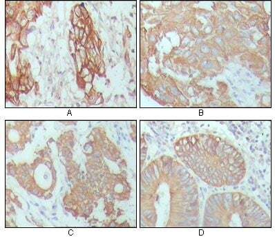
- Immunohistochemical analysis of paraffin-embedded human breast carcinoma (A), hepatocarcinoma (B), stomach cancer (C) and colon cancer tissue (D), showing cytoplasmic location using Cytokeratin 18 monoclonal antibody (Product # MA5-15458) followed with DAB staining