 Explore
 Validate
 Learn
Learn Western blot
 Immunocytochemistry
Antibody data
- Antibody Data
- Antigen structure
- References [3]
- Comments [0]
- Validations
- Immunocytochemistry [1]
Submit
Validation data
Reference
Comment
Report error
- Product number
- HPA001605 - Provider product page

- Provider
- Atlas Antibodies
- Proper citation
- Atlas Antibodies Cat#HPA001605, RRID:AB_2666381
- Product name
- Anti-KRT18
- Antibody type
- Polyclonal
- Description
- Polyclonal Antibody against Human KRT18, Gene description: keratin 18, Validated applications: ICC, IHC, WB, Uniprot ID: P05783, Storage: Store at +4°C for short term storage. Long time storage is recommended at -20°C.
- Reactivity
- Human
- Host
- Rabbit
- Conjugate
- Unconjugated
- Isotype
- IgG
- Vial size
- 100 µl
- Concentration
- 0.1 mg/ml
- Storage
- Store at +4°C for short term storage. Long time storage is recommended at -20°C.
- Handling
- The antibody solution should be gently mixed before use.
Submitted references An alternative splicing signature defines the basal-like phenotype and predicts worse clinical outcome in pancreatic cancer
Circulating Tumour Cells Indicate the Presence of Residual Disease Post-Castration in Prostate Cancer Patient-Derived Xenograft Models
Diversity of Epithelial-Mesenchymal Phenotypes in Circulating Tumour Cells from Prostate Cancer Patient-Derived Xenograft Models
Ruta V, Naro C, Pieraccioli M, Leccese A, Archibugi L, Cesari E, Panzeri V, Allgöwer C, Arcidiacono P, Falconi M, Carbone C, Tortora G, Borrelli F, Attili F, Spada C, Quero G, Alfieri S, Doglioni C, Kleger A, Capurso G, Sette C
Cell Reports Medicine 2024;5(2):101411
Cell Reports Medicine 2024;5(2):101411
Circulating Tumour Cells Indicate the Presence of Residual Disease Post-Castration in Prostate Cancer Patient-Derived Xenograft Models
Hassan S, Blick T, Wood J, Thompson E, Williams E
Frontiers in Cell and Developmental Biology 2022;10
Frontiers in Cell and Developmental Biology 2022;10
Diversity of Epithelial-Mesenchymal Phenotypes in Circulating Tumour Cells from Prostate Cancer Patient-Derived Xenograft Models
Hassan S, Blick T, Thompson E, Williams E
Cancers 2021;13(11):2750
Cancers 2021;13(11):2750
No comments: Submit comment
Supportive validation
- Submitted by
- Atlas Antibodies (provider)
- Main image
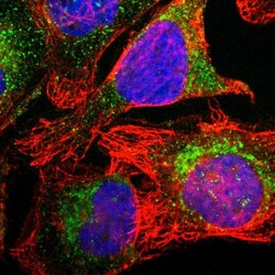
- Experimental details
- Immunofluorescent staining of human cell line U-2 OS shows localization to cytosol.
- Sample type
- Human
