 Explore
 Validate
 Learn
Learn Western blot
 Flow cytometry
Antibody data
- Antibody Data
- Antigen structure
- References [2]
- Comments [0]
- Validations
- Western blot [2]
- Immunocytochemistry [2]
- Immunohistochemistry [9]
- Other assay [1]
Submit
Validation data
Reference
Comment
Report error
- Product number
- MA1-06326 - Provider product page

- Provider
- Invitrogen Antibodies
- Product name
- Cytokeratin 18 Monoclonal Antibody (RGE53)
- Antibody type
- Monoclonal
- Antigen
- Other
- Description
- MA1-06326 detects cytokeratin 18 in human, mouse, rat, hamster, rabbit, chicken, canine, zebrafish and swine samples. MA1-06326 has sucessfully been used in Western blotting, immunocytochemistry, flow cytometry, and immunohistochemistry. The MA1-06326 immunogen is a cytoskeletal preparation from cells. Store at 4ºC or in small aliquots at -20ºC.
- Reactivity
- Human, Mouse, Rat, Canine, Chicken/Avian, Hamster, Porcine, Rabbit, Zebrafish
- Host
- Mouse
- Isotype
- IgG
- Antibody clone number
- RGE53
- Vial size
- 100 µL
- Concentration
- 1 mg/mL
- Storage
- Store at 4°C short term. For long term storage, store at -20°C, avoiding freeze/thaw cycles.
Submitted references Uptake of Manganese from the Manganese-Lysine Complex in Primary Chicken Intestinal Epithelial Cells.
Polyethylene glycol (PEG)-induced mouse model of choroidal neovascularization.
Bai S, Zhang K, Ding X, Wang J, Zeng Q, Peng H, Bai J, Xuan Y, Su Z, Wu B
Animals : an open access journal from MDPI 2019 Aug 15;9(8)
Animals : an open access journal from MDPI 2019 Aug 15;9(8)
Polyethylene glycol (PEG)-induced mouse model of choroidal neovascularization.
Lyzogubov VV, Tytarenko RG, Liu J, Bora NS, Bora PS
The Journal of biological chemistry 2011 May 6;286(18):16229-37
The Journal of biological chemistry 2011 May 6;286(18):16229-37
No comments: Submit comment
Supportive validation
- Submitted by
- Invitrogen Antibodies (provider)
- Main image
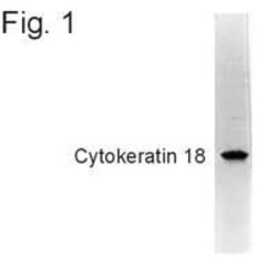
- Experimental details
- Western blot detection of cytokeratin 18 in cytoskeletal preparations from bovine lens tissue using Product # MA1-06326.

- Submitted by
- Invitrogen Antibodies (provider)
- Main image
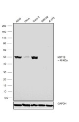
- Experimental details
- Western blot was performed using Anti-Cytokeratin 18 Monoclonal Antibody (RGE53)(Product # MA1-06326) and a 48kDa band corresponding to Cytokeratin 18 was observed across the cell lines tested except IMR-32 and A-375. Whole cell extracts (30 µg lysate) of A549 (Lane 1), HeLa (Lane 2), Caco-2 (Lane 3), IMR-32 (Lane 4) and A-375 (Lane 5) were electrophoresed using NuPAGE™ 4-12% Bis-Tris Protein Gel (Product # NP0321BOX). Resolved proteins were then transferred onto a Nitrocellulose membrane (Product # LC2001) by iBlot® 2 Dry Blotting System (Product # IB21001). The blot was probed with the primary antibody (1:1000 dilution) and detected by chemiluminescence with Goat anti-Mouse IgG (H+L) Superclonal™ Recombinant Secondary Antibody, HRP (Product # A28177,1:4000 dilution) using the iBright FL 1000 (Product # A32752). Chemiluminescent detection was performed using Novex® ECL Chemiluminescent Substrate Reagent Kit (Product # WP20005).

Supportive validation
- Submitted by
- Invitrogen Antibodies (provider)
- Main image
- Experimental details
- Immunostaining of Cytokeratin 18 in the glandular part of the human cervix using Product # MA1-06326.

- Submitted by
- Invitrogen Antibodies (provider)
- Main image

- Experimental details
- Immunofluorescence analysis of Cytokeratin 18 was performed using 70% confluent log phase Caco-2 cells. The cells were fixed with ice-cold acetone at 4°C for 5 minutes and blocked with 2% BSA for 45 minutes at room temperature. The cells were labeled with Cytokeratin 18 Monoclonal Antibody (RGE53) (Product # MA1-06326) at 1:100 dilution in 0.1% BSA, incubated at 4 degree celsius overnight and then labeled with Goat anti-Mouse IgG (H+L) Superclonal™ Recombinant Secondary Antibody, Alexa Fluor® 488 conjugate (Product # A28175), (1:2000 dilution), for 45 minutes at room temperature (Panel a: Green). Nuclei (Panel b: Blue) were stained with SlowFade® Gold Antifade Mountant with DAPI (Product # S36938). F-actin (Panel c: Red) was stained with Rhodamine Phalloidin (Product # R415, 1:300). Panel d represents the merged image showing cytoskeletal localization. Panel e represents A-375 cells which lack cytokeratin 18 expression. Panel f represents control cells with no primary antibody to assess background. The images were captured at 60X magnification.

Supportive validation
- Submitted by
- Invitrogen Antibodies (provider)
- Main image
- Experimental details
- Immunofluorescent analysis of 5 days old zebrafish embryo. using Cytokeratin 18 monoclonal antibody (Product # MA1-06326). Left panel: DAPI-staining of cell nuclei, providing an overview of the tissue section used for immunostaining

- Submitted by
- Invitrogen Antibodies (provider)
- Main image
- Experimental details
- Immunofluorescent analysis of 1 month old zebrafish embryo using Cytokeratin 18 monoclonal antibody (Product # MA1-06326).

- Submitted by
- Invitrogen Antibodies (provider)
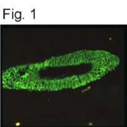
- Main image
- Experimental details
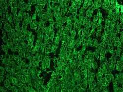
- Immunofluorescent analysis of epidermis of 2 days old zebrafish embryo using Cytokeratin 18 monoclonal antibody (Product # MA1-06326).

- Submitted by
- Invitrogen Antibodies (provider)
- Main image

- Experimental details
- Immunofluorescent analysis of epidermis of 2 days old zebrafish embryo using Cytokeratin 18 monoclonal antibody (Product # MA1-06326).

- Submitted by
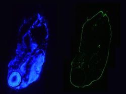
- Invitrogen Antibodies (provider)
- Main image

- Experimental details
- Immunofluorescent analysis of 5 days old zebrafish embryo. using Cytokeratin 18 monoclonal antibody (Product # MA1-06326). Left panel: DAPI-staining of cell nuclei, providing an overview of the tissue section used for immunostaining

- Submitted by
- Invitrogen Antibodies (provider)
- Main image

- Experimental details
- Immunofluorescent analysis of 1 month old zebrafish embryo using Cytokeratin 18 monoclonal antibody (Product # MA1-06326).

- Submitted by
- Invitrogen Antibodies (provider)
- Main image
- Experimental details
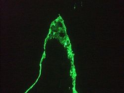
- Immunohistochemistry on frozen section of human colon stained with Cytokeratin 18 monoclonal antibody (Product # MA1-06326).

- Submitted by
- Invitrogen Antibodies (provider)
- Main image

- Experimental details
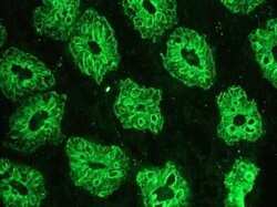
- Immunohistochemistry on frozen section of human kidney epithelium stained with Cytokeratin 18 monoclonal antibody (Product # MA1-06326).

- Submitted by
- Invitrogen Antibodies (provider)
- Main image
- Experimental details
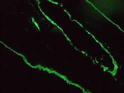
- Immunohistochemistry on frozen section of swine liver hepatocytes stained with Cytokeratin 18 monoclonal antibody (Product # MA1-06326).

Supportive validation
- Submitted by
- Invitrogen Antibodies (provider)
- Main image

- Experimental details
- Figure 1 Morphological features and immunofluorescence staining of primary chicken intestinal epithelial cells (IECs). ( A ) Epithelial cells were isolated in cell clusters. ( B ) Primary chicken IECs at 24 h post-seeding. The image shows cobblestone-shaped IECs radially growing from the center of the crypt (Cr) and cell clusters connecting. ( C ) Primary chicken IECs culture within 4-5 days post-seeding. Image shows a confluent chicken IECs. ( D , G , J ) Nuclei were stained (blue). ( E ) Immunostaining against cytokeratin-18 (green) at 3 days post-seeding. ( F ) Immunostaining against vimentin (red, the arrows in the figure) at 3 days post-seeding. ( H ) Primary chicken IECs presented in the image ( G ) were incubated only with the primary antibody of cytokeratin without the secondary antibody to serve as a primary antibody control. ( I ) Primary chicken IECs presented in the image ( G ) were incubated first with buffer only without the primary antibody of cytokeratin-18 and subsequently with secondary antibody to serve as a secondary antibody control. ( K ) Primary chicken IECs presented in the image ( J ) were incubated only with the primary antibody of cytokeratin without the secondary antibody to serve as a primary antibody control. ( L ) Primary chicken IECs presented in the image ( J ) were incubated first with buffer only without the primary antibody of cytokeratin-18 and subsequently with secondary antibody to serve as a secondary antibody control.
