Explore
 Validate
 Learn
Learn Western blot
Antibody data
- Antibody Data
- Antigen structure
- References [0]
- Comments [0]
- Validations
- Western blot [1]
- Immunocytochemistry [2]
- Immunohistochemistry [3]
Submit
Validation data
Reference
Comment
Report error
- Product number
- MA1-06327 - Provider product page

- Provider
- Invitrogen Antibodies
- Product name
- Cytokeratin 18 Monoclonal Antibody (RCK106)
- Antibody type
- Monoclonal
- Antigen
- Other
- Description
- MA1-06327 detects cytokeratin 18 in human samples.
- Antibody clone number
- RCK106
- Concentration
- 1 mg/mL
No comments: Submit comment
Supportive validation
- Submitted by
- Invitrogen Antibodies (provider)
- Main image
- Experimental details
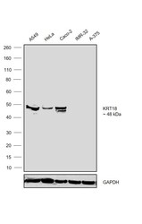
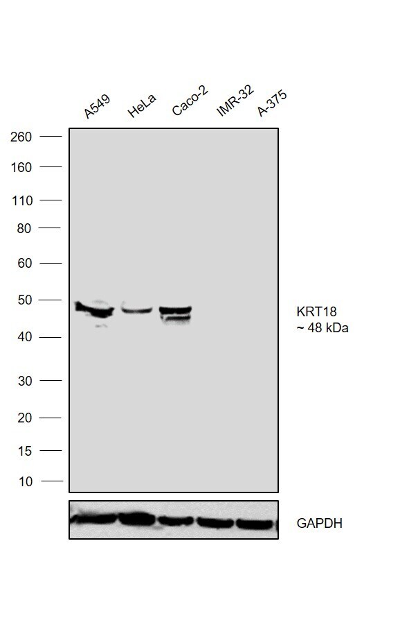
- Western blot was performed using Anti-Cytokeratin 18 Monoclonal Antibody (RCK106)(Product # MA1-06327) and a 48kDa band corresponding to Cytokeratin 18 was observed across the cell lines tested except IMR-32 and A-375. Whole cell extracts (30 µg lysate) of A549 (Lane 1), HeLa (Lane 2), Caco-2 (Lane 3), IMR-32 (Lane 4) and A-375 (Lane 5) were electrophoresed using NuPAGE™ 4-12% Bis-Tris Protein Gel (Product # NP0321BOX). Resolved proteins were then transferred onto a Nitrocellulose membrane (Product # LC2001) by iBlot® 2 Dry Blotting System (Product # IB21001). The blot was probed with the primary antibody (1:1000 dilution) and detected by chemiluminescence with Goat anti-Mouse IgG (H+L) Superclonal™ Recombinant Secondary Antibody, HRP (Product # A28177,1:4000 dilution) using the iBright FL 1000 (Product # A32752). Chemiluminescent detection was performed using Novex® ECL Chemiluminescent Substrate Reagent Kit (Product # WP20005).
Supportive validation
- Submitted by
- Invitrogen Antibodies (provider)
- Main image

- Experimental details
- Immunostaining of cytokeratin 18 in HaCat cells, a human keratinocyte culture, using Product # MA1-06327.
- Submitted by
- Invitrogen Antibodies (provider)
- Main image

- Experimental details
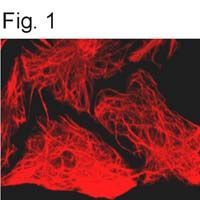
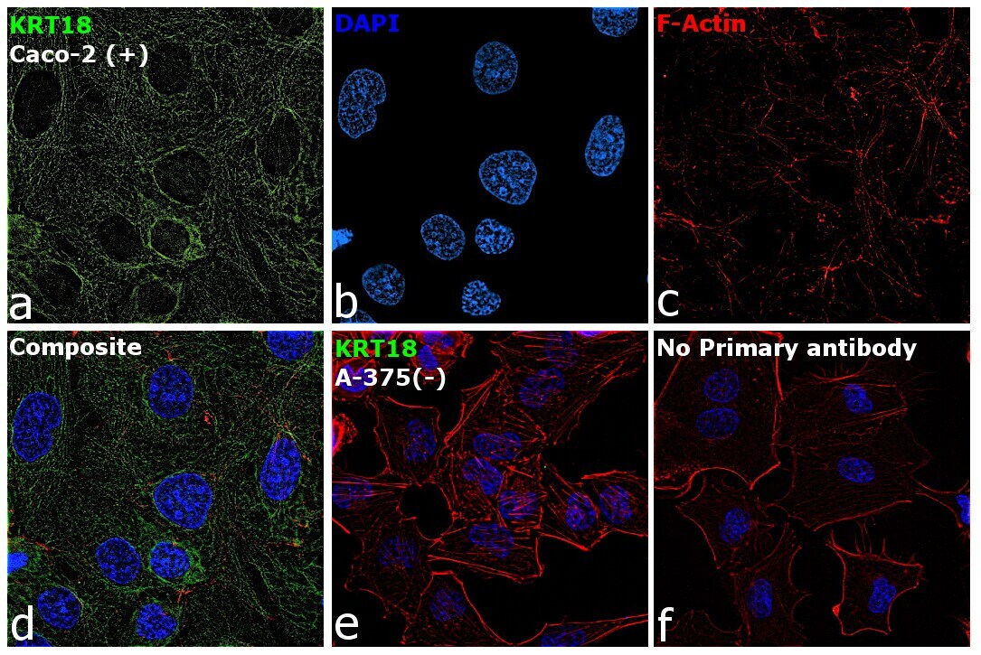
- Immunofluorescence analysis of Cytokeratin 18 was performed using 70% confluent log phase Caco-2 cells. The cells were fixed with ice-cold acetone at 4°C for 5 minutes and blocked with 2% BSA for 45 minutes at room temperature. The cells were labeled with Cytokeratin 18 Monoclonal Antibody (RCK106) (Product # MA1-06327) at 1:100 dilution in 0.1% BSA, incubated at 4 degree celsius overnight and then labeled with Goat anti-Mouse IgG (H+L) Superclonal™ Recombinant Secondary Antibody, Alexa Fluor® 488 conjugate (Product # A28175), (1:2000 dilution), for 45 minutes at room temperature (Panel a: Green). Nuclei (Panel b: Blue) were stained with SlowFade® Gold Antifade Mountant with DAPI (Product # S36938). F-actin (Panel c: Red) was stained with Rhodamine Phalloidin (Product # R415, 1:300 dilution). Panel d represents the merged image showing cytoskeletal localization. Panel e represents A-375 cells lacking cytoskeletal staining. Panel f represents control cells with no primary antibody to assess background. The images were captured at 60X magnification.
Supportive validation
- Submitted by
- Invitrogen Antibodies (provider)
- Main image
- Experimental details
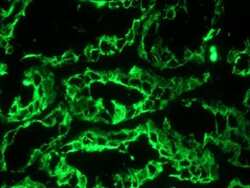
- Immunohistochemistry on frozen section of human kidney stained with Cytokeratin 18 monoclonal antibody (Product # MA1-06327).

- Submitted by
- Invitrogen Antibodies (provider)
- Main image

- Experimental details
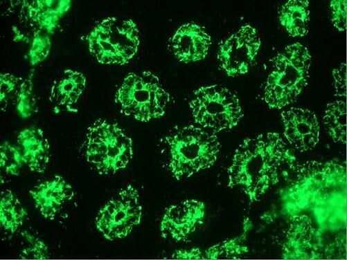
- Immunohistochemistry on frozen section of human small intestine stained with Cytokeratin 18 monoclonal antibody (Product # MA1-06327).
- Submitted by
- Invitrogen Antibodies (provider)
- Main image
- Experimental details
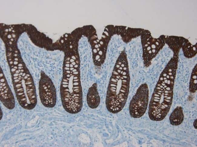
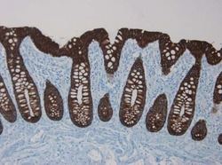
- Immunohistochemistry on paraffin section of human colon stained with Cytokeratin 18 monoclonal antibody (Product # MA1-06327).