 Explore
 Validate
 Learn
Learn Western blot
Antibody data
- Antibody Data
- Antigen structure
- References [2]
- Comments [0]
- Validations
- Western blot [2]
- Immunocytochemistry [2]
- Immunohistochemistry [3]
- Flow cytometry [1]
- Other assay [2]
Submit
Validation data
Reference
Comment
Report error
- Product number
- PA5-14263 - Provider product page

- Provider
- Invitrogen Antibodies
- Product name
- Cytokeratin 18 Polyclonal Antibody
- Antibody type
- Polyclonal
- Antigen
- Synthetic peptide
- Reactivity
- Human, Mouse
- Host
- Rabbit
- Isotype
- IgG
- Vial size
- 400 µL
- Concentration
- 0.5 mg/mL
- Storage
- Store at 4°C short term. For long term storage, store at -20°C, avoiding freeze/thaw cycles.
Submitted references YAP1/TAZ drives ependymoma-like tumour formation in mice.
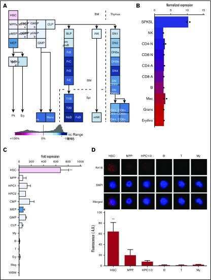
Lineage tracing of murine adult hematopoietic stem cells reveals active contribution to steady-state hematopoiesis.
Eder N, Roncaroli F, Domart MC, Horswell S, Andreiuolo F, Flynn HR, Lopes AT, Claxton S, Kilday JP, Collinson L, Mao JH, Pietsch T, Thompson B, Snijders AP, Ultanir SK
Nature communications 2020 May 13;11(1):2380
Nature communications 2020 May 13;11(1):2380
Lineage tracing of murine adult hematopoietic stem cells reveals active contribution to steady-state hematopoiesis.
Chapple RH, Tseng YJ, Hu T, Kitano A, Takeichi M, Hoegenauer KA, Nakada D
Blood advances 2018 Jun 12;2(11):1220-1228
Blood advances 2018 Jun 12;2(11):1220-1228
No comments: Submit comment
Supportive validation
- Submitted by
- Invitrogen Antibodies (provider)
- Main image

- Experimental details
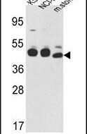
- Western blot analysis using a CYK18 polyclonal antibody (Product # PA5-14263) in K562, NCI-H460 cell lysates and mouse stomach tissues lysates (35 µg per lane).
- Submitted by
- Invitrogen Antibodies (provider)
- Main image

- Experimental details
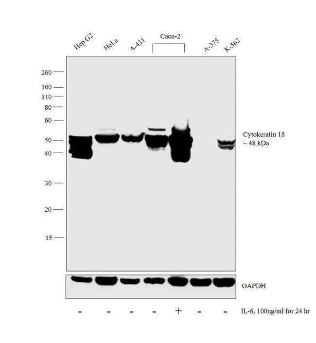
- Western blot analysis was performed on whole cell extracts (30 µg lysate) of Hep G2 (Lane 1), HeLa (Lane 2), A-431 (Lane 3), Caco-2 (Lane 4) Caco-2 treated with IL-6 (100ng/ml for 24hr) (Lane 5), A-375 (Lane 6) and K-562 (Lane 7). The blot was probed with Anti-Cytokeratin 18 Polyclonal Antibody (Product # PA5-14263, 1:1000 dilution) and detected by chemiluminescence using Goat anti-Rabbit IgG (H+L) Superclonal™ Secondary Antibody, HRP conjugate (Product # A27036, 0.25 µg/ml, 1:4000 dilution). A 48 kDa band corresponding to Cytokeratin 18 was observed across the cell lines tested. Also, the protein expression increased in Caco-2 cell line upon IL-6 treatment and was not detected in A-375 which is reported to be negative for Cytokeratin 18.
Supportive validation
- Submitted by
- Invitrogen Antibodies (provider)
- Main image

- Experimental details
- Immunofluorescent analysis of HeLa cells using a CYK18 polyclonal antibody (Product # PA5-14263) at a dilution of 1:10-50, followed by a fluor-conjugated goat anti-rabbit secondary antibody (green). Actin filaments were stained with dye-conjugated phalloidin (red). Nuclei were stained with DAPI (blue).

- Submitted by
- Invitrogen Antibodies (provider)
- Main image

- Experimental details
- Immunofluorescence analysis of Cytokeratin 18 was performed using 70% confluent log phase Hep G2 cells. The cells were fixed and permeabilized with ice-cold acetone and blocked with 1% BSA for 1 hour at room temperature. The cells were labeled with Cytokeratin 18 Polyclonal Antibody (Product # PA5-14263) at 1:50 dilution in 0.1% BSA, incubated at 4 degree Celsius overnight and then labeled with Goat anti-Rabbit IgG (H+L) Superclonal™ Secondary Antibody, Alexa Fluor® 488 conjugate (Product # A27034) at a dilution of 1:2000 for 45 minutes at room temperature (Panel a: green). Nuclei (Panel b: blue) were stained with ProLong™ Diamond Antifade Mountant with DAPI (Product # P36962). F-actin (Panel c: red) was stained with Rhodamine Phalloidin (Product # R415). Panel d represents the merged image showing cytoskeletal localization. Panel e represents control cells with no primary antibody to assess background. Panel f shows the negative cell line A-375 with no Cytokeratin 18 expression. The images were captured at 60X magnification.

Supportive validation
- Submitted by
- Invitrogen Antibodies (provider)
- Main image

- Experimental details
- Immunohistochemical analysis of formalin-fixed, paraffin-embedded human colon carcinoma tissue using a CYK18 polyclonal antibody (Product # PA5-14263), followed by HRP-conjugated secondary antibody and DAB staining.

- Submitted by
- Invitrogen Antibodies (provider)
- Main image

- Experimental details
- Immunofluorescent analysis of prostate carcinoma using a CYK18 polyclonal antibody (Product # PA5-14263) at a dilution of 1:10-50, followed by a fluor-conjugated goat anti-rabbit secondary antibody (green). Nuclei were stained with DAPI (blue).

- Submitted by
- Invitrogen Antibodies (provider)
- Main image

- Experimental details
- Immunohistochemistry analysis in formalin-fixed, paraffin-embedded human prostate carcinoma using a CYK18 polyclonal antibody (Product # PA5-14263), followed by HRP-conjugated secondary antibody and DAB staining.

Supportive validation
- Submitted by
- Invitrogen Antibodies (provider)
- Main image

- Experimental details
- Flow cytometry analysis of WiDr cells using a CYK18 polyclonal antibody (Product # PA5-14263) (bottom), compared to a negative control cell (top) at a dilution of 1:10-50, followed by a FITC-conjugated goat anti-rabbit antibody

Supportive validation
- Submitted by
- Invitrogen Antibodies (provider)
- Main image
- Experimental details
- NULL
- Submitted by
- Invitrogen Antibodies (provider)
- Main image

- Experimental details
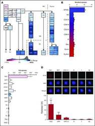
- Fig. 5 LATS1/2 cKO mouse tumours display histological features of ependymoma. a - d The tumour (arrowhead) appear to arise from the ependymal layer of the anterior temporal horn of the lateral ventricle at the level of hippocampus (* CA3). a H&E, x4, scale bar = 500 um; the lesion has compressive margin. b H&E, x10, scale bar = 200 um and it is composed of fascicles of spindle cells with focal nuclear clustering. c . H&E, x40, scale bar = 50 um. A few tumour cells contain intracytoplasmic, paranuclear vacuoles (arrowheads). d . H&E, x60, scale bar = 20 um. Tumour cells demonstrate. e nuclear and cytoplasmic YAP1 expression (immunoperoxidase, x20), f weak cytoplasmic MUC1 expression (immunoperoxidase, x20) and g they are focally positive for GFAP (immunoperoxidase, x20) and h cytokeratin 18/CK18 (immunoperoxidase, x10). Reactive astrocytes surrounding the lesion are also GFAP-positive ( g --immunoperoxidase, x20). Scale bars = 50 um. i TEM image (x9900) within the tumour showing multiple ependymal features (Yellow arrowheads = microvilli, red arrows = tight junctions). Scale bar = 1 um. j , k . Enlargement of the yellow and red insets in i , respectively. Scale bars = 200 nm. j . High magnification of microvilli and k . a tight junction found in the tumour are shown.
