 Explore
 Validate
 Learn
Learn Western blot
 Immunocytochemistry
Antibody data
- Antibody Data
- Antigen structure
- References [46]
- Comments [0]
- Validations
- Immunocytochemistry [2]
- Immunohistochemistry [1]
- Flow cytometry [1]
- Other assay [8]
Submit
Validation data
Reference
Comment
Report error
- Product number
- MA5-12104 - Provider product page

- Provider
- Invitrogen Antibodies
- Product name
- Cytokeratin 18 Monoclonal Antibody (DC10)
- Antibody type
- Monoclonal
- Antigen
- Other
- Description
- MA5-12104 targets Cytokeratin 18 in IF, IHC (P), FACS, and WB applications and shows reactivity with Human samples. The MA5-12104 immunogen is human breast cancer PMC 42 cells.
- Reactivity
- Human, Mouse
- Host
- Mouse
- Isotype
- IgG
- Antibody clone number
- DC10
- Vial size
- 500 μL
- Concentration
- 0.2 mg/mL
- Storage
- 4°C
Submitted references CRISPR interference interrogation of COPD GWAS genes reveals the functional significance of desmoplakin in iPSC-derived alveolar epithelial cells.
Cis-regulatory chromatin loops analysis identifies GRHL3 as a master regulator of surface epithelium commitment.
Primary ectocervical epithelial cells display lower permissivity to Chlamydia trachomatis than HeLa cells and a globally higher pro-inflammatory profile.
Interferon-τ regulates the expression and function of bovine leukocyte antigen by downregulating bta-miR-204.
Survival Pathways Are Differently Affected by Microgravity in Normal and Cancerous Breast Cells.
Applicability of Artificial Vascularized Liver Tissue to Proteomic Analysis.
The Coxiella burnetii T4SS Effector AnkF Is Important for Intracellular Replication.
Engineered Liver Tissue Culture in an In Vitro Tubular Perfusion System.
Mechanisms of Endogenous HIV-1 Reactivation by Endocervical Epithelial Cells.
Downregulation of C-Terminal Tensin-Like Protein (CTEN) Suppresses Prostate Cell Proliferation and Contributes to Acinar Morphogenesis.
Casein kinase 1 is recruited to nuclear speckles by FAM83H and SON.
Vitamin D and androgen receptor-targeted therapy for triple-negative breast cancer.
FAM83H and casein kinase I regulate the organization of the keratin cytoskeleton and formation of desmosomes.
Ambient Light Promotes Selective Subcellular Proteotoxicity after Endogenous and Exogenous Porphyrinogenic Stress.
Modelling breast cancer requires identification and correction of a critical cell lineage-dependent transduction bias.
Alterations of canalicular ATP-binding cassette transporter expression in drug-induced liver injury.
A novel mechanism of keratin cytoskeleton organization through casein kinase Iα and FAM83H in colorectal cancer.
Glucose and SIRT2 reciprocally mediate the regulation of keratin 8 by lysine acetylation.
Malignant transformation of adenomyoepithelioma of the breast by a monophasic population: a report of two cases and review of literature.
Expression of KRT7 and WT1 differentiates precursor lesions of Wilms' tumours from those of papillary renal cell tumours and mucinous tubular and spindle cell carcinomas.
Keratin hypersumoylation alters filament dynamics and is a marker for human liver disease and keratin mutation.
Directed differentiation of human embryonic stem cells to epidermal progenitors.
Giant clear cell hidradenoma of the knee.
Cytoskeletal keratin glycosylation protects epithelial tissue from injury.
Antagonistic roles of Notch and p63 in controlling mammary epithelial cell fates.
Endometrial profile of tamoxifen and low-dose estradiol combination therapy.
Imaging and analysis of 3D tumor spheroids enriched for a cancer stem cell phenotype.
Assessment of the tumorigenesis and drug susceptibility of three new canine mammary tumor cell lines.
Antigen expression of human eccrine sweat glands.
Long-term cultures of bone marrow-derived human mesenchymal stem cells frequently undergo spontaneous malignant transformation.
Frzb, a secreted Wnt antagonist, decreases growth and invasiveness of fibrosarcoma cells associated with inhibition of Met signaling.
Monitoring of epithelial cell caspase activation via detection of durable keratin fragment formation.
An immunohistochemical profile of giant cell carcinoma of the larynx.
Mammary gland development in early pubertal female macaques.
Cellular and molecular mechanisms of abnormal calcification following ischemia-reperfusion injury in human liver transplantation.
Analysis of keratin polypeptides 8 and 19 variants in inflammatory bowel disease.
Blocking Wnt/LRP5 signaling by a soluble receptor modulates the epithelial to mesenchymal transition and suppresses met and metalloproteinases in osteosarcoma Saos-2 cells.
Identification of 14-3-3sigma as a contributor to drug resistance in human breast cancer cells using functional proteomic analysis.
Bispecific and human disease-related anti-keratin rabbit monoclonal antibodies.
Protein phosphatase-2A associates with and dephosphorylates keratin 8 after hyposmotic stress in a site- and cell-specific manner.
Expression of Frzb/secreted Frizzled-related protein 3, a secreted Wnt antagonist, in human androgen-independent prostate cancer PC-3 cells suppresses tumor growth and cellular invasiveness.
Studying simple epithelial keratins in cells and tissues.
Cytokeratin expression in lichen amyloidosus and macular amyloidosis.
Keratin 8 and 18 hyperphosphorylation is a marker of progression of human liver disease.
Keratin mutation in transgenic mice predisposes to Fas but not TNF-induced apoptosis and massive liver injury.
Keratin 8 mutations in patients with cryptogenic liver disease.
Werder RB, Liu T, Abo KM, Lindstrom-Vautrin J, Villacorta-Martin C, Huang J, Hinds A, Boyer N, Bullitt E, Liesa M, Silverman EK, Kotton DN, Cho MH, Zhou X, Wilson AA
Science advances 2022 Jul 15;8(28):eabo6566
Science advances 2022 Jul 15;8(28):eabo6566
Cis-regulatory chromatin loops analysis identifies GRHL3 as a master regulator of surface epithelium commitment.
Huang H, Liu J, Li M, Guo H, Zhu J, Zhu L, Wu S, Mo K, Huang Y, Tan J, Chen C, Wang B, Yu Y, Wang L, Liu Y, Ouyang H
Science advances 2022 Jul 15;8(28):eabo5668
Science advances 2022 Jul 15;8(28):eabo5668
Primary ectocervical epithelial cells display lower permissivity to Chlamydia trachomatis than HeLa cells and a globally higher pro-inflammatory profile.
Tang C, Liu C, Maffei B, Niragire B, Cohen H, Kane A, Donnadieu AC, Levy-Zauberman Y, Vernay T, Hugueny J, Vincens E, Louis-Sylvestre C, Subtil A, Wu Y
Scientific reports 2021 Mar 12;11(1):5848
Scientific reports 2021 Mar 12;11(1):5848
Interferon-τ regulates the expression and function of bovine leukocyte antigen by downregulating bta-miR-204.
Wang X, Yuan T, Yin N, Ma X, Yang Y, Yang J, Shaukat A, Deng G
Experimental and therapeutic medicine 2021 Jun;21(6):594
Experimental and therapeutic medicine 2021 Jun;21(6):594
Survival Pathways Are Differently Affected by Microgravity in Normal and Cancerous Breast Cells.
Monti N, Masiello MG, Proietti S, Catizone A, Ricci G, Harrath AH, Alwasel SH, Cucina A, Bizzarri M
International journal of molecular sciences 2021 Jan 16;22(2)
International journal of molecular sciences 2021 Jan 16;22(2)
Applicability of Artificial Vascularized Liver Tissue to Proteomic Analysis.
Mori N, Kida YS
Micromachines 2021 Apr 11;12(4)
Micromachines 2021 Apr 11;12(4)
The Coxiella burnetii T4SS Effector AnkF Is Important for Intracellular Replication.
Pechstein J, Schulze-Luehrmann J, Bisle S, Cantet F, Beare PA, Ölke M, Bonazzi M, Berens C, Lührmann A
Frontiers in cellular and infection microbiology 2020;10:559915
Frontiers in cellular and infection microbiology 2020;10:559915
Engineered Liver Tissue Culture in an In Vitro Tubular Perfusion System.
Yang G, Mahadik B, Mollot T, Pinsky J, Jones A, Robinson A, Najafali D, Rivkin D, Katsnelson J, Piard C, Fisher JP
Tissue engineering. Part A 2020 Dec;26(23-24):1369-1377
Tissue engineering. Part A 2020 Dec;26(23-24):1369-1377
Mechanisms of Endogenous HIV-1 Reactivation by Endocervical Epithelial Cells.
Gornalusse GG, Valdez R, Fenkart G, Vojtech L, Fleming LM, Pandey U, Hughes SM, Levy CN, Dela Cruz EJ, Calienes FL, Kirby AC, Fialkow MF, Lentz GM, Wagoner J, Jing L, Koelle DM, Polyak SJ, Fredricks DN, McElrath MJ, Wald A, Hladik F
Journal of virology 2020 Apr 16;94(9)
Journal of virology 2020 Apr 16;94(9)
Downregulation of C-Terminal Tensin-Like Protein (CTEN) Suppresses Prostate Cell Proliferation and Contributes to Acinar Morphogenesis.
Wu WM, Liao YC
International journal of molecular sciences 2018 Oct 16;19(10)
International journal of molecular sciences 2018 Oct 16;19(10)
Casein kinase 1 is recruited to nuclear speckles by FAM83H and SON.
Kuga T, Kume H, Adachi J, Kawasaki N, Shimizu M, Hoshino I, Matsubara H, Saito Y, Nakayama Y, Tomonaga T
Scientific reports 2016 Sep 29;6:34472
Scientific reports 2016 Sep 29;6:34472
Vitamin D and androgen receptor-targeted therapy for triple-negative breast cancer.
Thakkar A, Wang B, Picon-Ruiz M, Buchwald P, Ince TA
Breast cancer research and treatment 2016 May;157(1):77-90
Breast cancer research and treatment 2016 May;157(1):77-90
FAM83H and casein kinase I regulate the organization of the keratin cytoskeleton and formation of desmosomes.
Kuga T, Sasaki M, Mikami T, Miake Y, Adachi J, Shimizu M, Saito Y, Koura M, Takeda Y, Matsuda J, Tomonaga T, Nakayama Y
Scientific reports 2016 May 25;6:26557
Scientific reports 2016 May 25;6:26557
Ambient Light Promotes Selective Subcellular Proteotoxicity after Endogenous and Exogenous Porphyrinogenic Stress.
Maitra D, Elenbaas JS, Whitesall SE, Basrur V, D'Alecy LG, Omary MB
The Journal of biological chemistry 2015 Sep 25;290(39):23711-24
The Journal of biological chemistry 2015 Sep 25;290(39):23711-24
Modelling breast cancer requires identification and correction of a critical cell lineage-dependent transduction bias.
Hines WC, Yaswen P, Bissell MJ
Nature communications 2015 Apr 21;6:6927
Nature communications 2015 Apr 21;6:6927
Alterations of canalicular ATP-binding cassette transporter expression in drug-induced liver injury.
Zollner G, Thueringer A, Lackner C, Fickert P, Trauner M
Digestion 2014;90(2):81-8
Digestion 2014;90(2):81-8
A novel mechanism of keratin cytoskeleton organization through casein kinase Iα and FAM83H in colorectal cancer.
Kuga T, Kume H, Kawasaki N, Sato M, Adachi J, Shiromizu T, Hoshino I, Nishimori T, Matsubara H, Tomonaga T
Journal of cell science 2013 Oct 15;126(Pt 20):4721-31
Journal of cell science 2013 Oct 15;126(Pt 20):4721-31
Glucose and SIRT2 reciprocally mediate the regulation of keratin 8 by lysine acetylation.
Snider NT, Leonard JM, Kwan R, Griggs NW, Rui L, Omary MB
The Journal of cell biology 2013 Feb 4;200(3):241-7
The Journal of cell biology 2013 Feb 4;200(3):241-7
Malignant transformation of adenomyoepithelioma of the breast by a monophasic population: a report of two cases and review of literature.
Marian C, Boila A, Soanca D, Malau M, Podeanu DM, Resetkova E, Stolnicu S
APMIS : acta pathologica, microbiologica, et immunologica Scandinavica 2013 Apr;121(4):272-9
APMIS : acta pathologica, microbiologica, et immunologica Scandinavica 2013 Apr;121(4):272-9
Expression of KRT7 and WT1 differentiates precursor lesions of Wilms' tumours from those of papillary renal cell tumours and mucinous tubular and spindle cell carcinomas.
Szponar A, Kovacs G
Virchows Archiv : an international journal of pathology 2012 Apr;460(4):423-7
Virchows Archiv : an international journal of pathology 2012 Apr;460(4):423-7
Keratin hypersumoylation alters filament dynamics and is a marker for human liver disease and keratin mutation.
Snider NT, Weerasinghe SV, Iñiguez-Lluhí JA, Herrmann H, Omary MB
The Journal of biological chemistry 2011 Jan 21;286(3):2273-84
The Journal of biological chemistry 2011 Jan 21;286(3):2273-84
Directed differentiation of human embryonic stem cells to epidermal progenitors.
Metallo CM, Ji L, de Pablo JJ, Palecek SP
Methods in molecular biology (Clifton, N.J.) 2010;585:83-92
Methods in molecular biology (Clifton, N.J.) 2010;585:83-92
Giant clear cell hidradenoma of the knee.
Yu G, Goodloe S Jr, D'Angelis CA, McGrath BE, Chen F
Journal of cutaneous pathology 2010 Sep;37(9):e37-41
Journal of cutaneous pathology 2010 Sep;37(9):e37-41
Cytoskeletal keratin glycosylation protects epithelial tissue from injury.
Ku NO, Toivola DM, Strnad P, Omary MB
Nature cell biology 2010 Sep;12(9):876-85
Nature cell biology 2010 Sep;12(9):876-85
Antagonistic roles of Notch and p63 in controlling mammary epithelial cell fates.
Yalcin-Ozuysal O, Fiche M, Guitierrez M, Wagner KU, Raffoul W, Brisken C
Cell death and differentiation 2010 Oct;17(10):1600-12
Cell death and differentiation 2010 Oct;17(10):1600-12
Endometrial profile of tamoxifen and low-dose estradiol combination therapy.
Wood CE, Kaplan JR, Fontenot MB, Williams JK, Cline JM
Clinical cancer research : an official journal of the American Association for Cancer Research 2010 Feb 1;16(3):946-56
Clinical cancer research : an official journal of the American Association for Cancer Research 2010 Feb 1;16(3):946-56
Imaging and analysis of 3D tumor spheroids enriched for a cancer stem cell phenotype.
Robertson FM, Ogasawara MA, Ye Z, Chu K, Pickei R, Debeb BG, Woodward WA, Hittelman WN, Cristofanilli M, Barsky SH
Journal of biomolecular screening 2010 Aug;15(7):820-9
Journal of biomolecular screening 2010 Aug;15(7):820-9
Assessment of the tumorigenesis and drug susceptibility of three new canine mammary tumor cell lines.
Chang CY, Chiou PP, Chen WJ, Li YH, Yiu JC, Cheng YH, Chen SD, Lin CT, Lai YS
Research in veterinary science 2010 Apr;88(2):285-93
Research in veterinary science 2010 Apr;88(2):285-93
Antigen expression of human eccrine sweat glands.
Li HH, Zhou G, Fu XB, Zhang L
Journal of cutaneous pathology 2009 Mar;36(3):318-24
Journal of cutaneous pathology 2009 Mar;36(3):318-24
Long-term cultures of bone marrow-derived human mesenchymal stem cells frequently undergo spontaneous malignant transformation.
Røsland GV, Svendsen A, Torsvik A, Sobala E, McCormack E, Immervoll H, Mysliwietz J, Tonn JC, Goldbrunner R, Lønning PE, Bjerkvig R, Schichor C
Cancer research 2009 Jul 1;69(13):5331-9
Cancer research 2009 Jul 1;69(13):5331-9
Frzb, a secreted Wnt antagonist, decreases growth and invasiveness of fibrosarcoma cells associated with inhibition of Met signaling.
Guo Y, Xie J, Rubin E, Tang YX, Lin F, Zi X, Hoang BH
Cancer research 2008 May 1;68(9):3350-60
Cancer research 2008 May 1;68(9):3350-60
Monitoring of epithelial cell caspase activation via detection of durable keratin fragment formation.
Tao GZ, Li DH, Zhou Q, Toivola DM, Strnad P, Sandesara N, Cheung RC, Hong A, Omary MB
The Journal of pathology 2008 Jun;215(2):164-74
The Journal of pathology 2008 Jun;215(2):164-74
An immunohistochemical profile of giant cell carcinoma of the larynx.
Gurbuz Y, Kose N, Aydin O, Ozturk M
Auris, nasus, larynx 2007 Sep;34(3):413-6
Auris, nasus, larynx 2007 Sep;34(3):413-6
Mammary gland development in early pubertal female macaques.
Wood CE, Hester JM, Cline JM
Toxicologic pathology 2007 Oct;35(6):795-805
Toxicologic pathology 2007 Oct;35(6):795-805
Cellular and molecular mechanisms of abnormal calcification following ischemia-reperfusion injury in human liver transplantation.
Kalantari F, Miao D, Emadali A, Tzimas GN, Goltzman D, Vali H, Chevet E, Auguste P
Modern pathology : an official journal of the United States and Canadian Academy of Pathology, Inc 2007 Mar;20(3):357-66
Modern pathology : an official journal of the United States and Canadian Academy of Pathology, Inc 2007 Mar;20(3):357-66
Analysis of keratin polypeptides 8 and 19 variants in inflammatory bowel disease.
Tao GZ, Strnad P, Zhou Q, Kamal A, Zhang L, Madani ND, Kugathasan S, Brant SR, Cho JH, Omary MB, Duerr RH
Clinical gastroenterology and hepatology : the official clinical practice journal of the American Gastroenterological Association 2007 Jul;5(7):857-64
Clinical gastroenterology and hepatology : the official clinical practice journal of the American Gastroenterological Association 2007 Jul;5(7):857-64
Blocking Wnt/LRP5 signaling by a soluble receptor modulates the epithelial to mesenchymal transition and suppresses met and metalloproteinases in osteosarcoma Saos-2 cells.
Guo Y, Zi X, Koontz Z, Kim A, Xie J, Gorlick R, Holcombe RF, Hoang BH
Journal of orthopaedic research : official publication of the Orthopaedic Research Society 2007 Jul;25(7):964-71
Journal of orthopaedic research : official publication of the Orthopaedic Research Society 2007 Jul;25(7):964-71
Identification of 14-3-3sigma as a contributor to drug resistance in human breast cancer cells using functional proteomic analysis.
Liu Y, Liu H, Han B, Zhang JT
Cancer research 2006 Mar 15;66(6):3248-55
Cancer research 2006 Mar 15;66(6):3248-55
Bispecific and human disease-related anti-keratin rabbit monoclonal antibodies.
Tao GZ, Nakamichi I, Ku NO, Wang J, Frolkis M, Gong X, Zhu W, Pytela R, Omary MB
Experimental cell research 2006 Feb 15;312(4):411-22
Experimental cell research 2006 Feb 15;312(4):411-22
Protein phosphatase-2A associates with and dephosphorylates keratin 8 after hyposmotic stress in a site- and cell-specific manner.
Tao GZ, Toivola DM, Zhou Q, Strnad P, Xu B, Michie SA, Omary MB
Journal of cell science 2006 Apr 1;119(Pt 7):1425-32
Journal of cell science 2006 Apr 1;119(Pt 7):1425-32
Expression of Frzb/secreted Frizzled-related protein 3, a secreted Wnt antagonist, in human androgen-independent prostate cancer PC-3 cells suppresses tumor growth and cellular invasiveness.
Zi X, Guo Y, Simoneau AR, Hope C, Xie J, Holcombe RF, Hoang BH
Cancer research 2005 Nov 1;65(21):9762-70
Cancer research 2005 Nov 1;65(21):9762-70
Studying simple epithelial keratins in cells and tissues.
Ku NO, Toivola DM, Zhou Q, Tao GZ, Zhong B, Omary MB
Methods in cell biology 2004;78:489-517
Methods in cell biology 2004;78:489-517
Cytokeratin expression in lichen amyloidosus and macular amyloidosis.
Apaydin R, Gürbüz Y, Bayramgürler D, Müezzinoglu B, Bilen N
Journal of the European Academy of Dermatology and Venereology : JEADV 2004 May;18(3):305-9
Journal of the European Academy of Dermatology and Venereology : JEADV 2004 May;18(3):305-9
Keratin 8 and 18 hyperphosphorylation is a marker of progression of human liver disease.
Toivola DM, Ku NO, Resurreccion EZ, Nelson DR, Wright TL, Omary MB
Hepatology (Baltimore, Md.) 2004 Aug;40(2):459-66
Hepatology (Baltimore, Md.) 2004 Aug;40(2):459-66
Keratin mutation in transgenic mice predisposes to Fas but not TNF-induced apoptosis and massive liver injury.
Ku NO, Soetikno RM, Omary MB
Hepatology (Baltimore, Md.) 2003 May;37(5):1006-14
Hepatology (Baltimore, Md.) 2003 May;37(5):1006-14
Keratin 8 mutations in patients with cryptogenic liver disease.
Ku NO, Gish R, Wright TL, Omary MB
The New England journal of medicine 2001 May 24;344(21):1580-7
The New England journal of medicine 2001 May 24;344(21):1580-7
No comments: Submit comment
Supportive validation
- Submitted by
- Invitrogen Antibodies (provider)
- Main image

- Experimental details
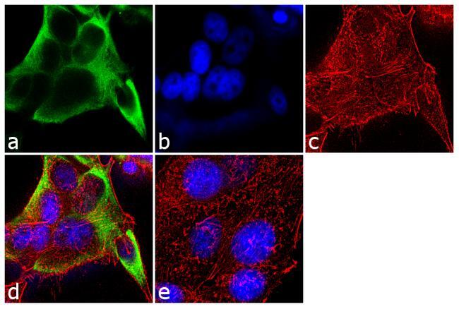
- Immunofluorescent analysis of Cytokeratin 18 was performed using 70% confluent log phase MCF-7 cells. The cells were fixed with 4% paraformaldehyde for 10 minutes, permeabilized with 0.1% Triton™ X-100 for 10 minutes, and blocked with 1% BSA for 1 hour at room temperature. The cells were labeled with Cytokeratin 18 (DC10) Mouse Monoclonal Antibody (Product # MA5-12104) at 2 µg/mL in 0.1% BSA and incubated for 3 hours at room temperature and then labeled with Goat anti-Mouse IgG (H+L) Superclonal™ Secondary Antibody, Alexa Fluor® 488 conjugate (Product # A28175) a dilution of 1:2000 for 45 minutes at room temperature (Panel a: green). Nuclei (Panel b: blue) were stained with SlowFade® Gold Antifade Mountant with DAPI (Product # S36938). F-actin (Panel c: red) was stained with Alexa Fluor® 555 Rhodamine Phalloidin (Product # R415, 1:300). Panel d represents the merged image showing cytoplasmic localization. Panel e shows the no primary antibody control. The images were captured at 60X magnification.

- Submitted by
- Invitrogen Antibodies (provider)
- Main image

- Experimental details
- Immunofluorescent analysis of Cytokeratin 18 was performed using 70% confluent log phase MCF-7 cells. The cells were fixed with 4% paraformaldehyde for 10 minutes, permeabilized with 0.1% Triton™ X-100 for 10 minutes, and blocked with 1% BSA for 1 hour at room temperature. The cells were labeled with Cytokeratin 18 (DC10) Mouse Monoclonal Antibody (Product # MA5-12104) at 2 µg/mL in 0.1% BSA and incubated for 3 hours at room temperature and then labeled with Goat anti-Mouse IgG (H+L) Superclonal™ Secondary Antibody, Alexa Fluor® 488 conjugate (Product # A28175) a dilution of 1:2000 for 45 minutes at room temperature (Panel a: green). Nuclei (Panel b: blue) were stained with SlowFade® Gold Antifade Mountant with DAPI (Product # S36938). F-actin (Panel c: red) was stained with Alexa Fluor® 555 Rhodamine Phalloidin (Product # R415, 1:300). Panel d represents the merged image showing cytoplasmic localization. Panel e shows the no primary antibody control. The images were captured at 60X magnification.
Supportive validation
- Submitted by
- Invitrogen Antibodies (provider)
- Main image
- Experimental details
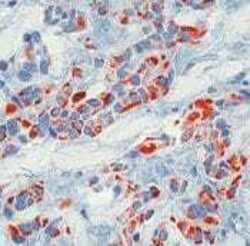
- Formalin-fixed, paraffin-embedded human breast cancer stained with Keratin 18 antibody using peroxidase-conjugate and AEC chromogen. Note cytoplasmic staining of tumor cells.

Supportive validation
- Submitted by
- Invitrogen Antibodies (provider)
- Main image
- Experimental details
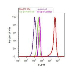
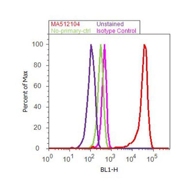
- Flow cytometry analysis of Cytokeratin 18 was done on MCF7 cells. Cells were fixed with 70% ethanol for 10 minutes, permeabilized with 0.25% Triton™ X-100 for 20 minutes, and blocked with 5% BSA for 30 minutes at room temperature. Cells were labeled with Cytokeratin 18 Mouse Monoclonal Antibody (MA5-12104, red histogram) or with mouse isotype control (pink histogram) at 3-5 ug/million cells in 2.5% BSA. After incubation at room temperature for 2 hours, the cells were labeled with Alexa Fluor® 488 Rabbit Anti-Mouse Secondary Antibody (A11059) at a dilution of 1:400 for 30 minutes at room temperature. The representative 10, 000 cells were acquired and analyzed for each sample using an Attune® Acoustic Focusing Cytometer. The purple histogram represents unstained control cells and the green histogram represents no-primary-antibody control.
Supportive validation
- Submitted by
- Invitrogen Antibodies (provider)
- Main image

- Experimental details
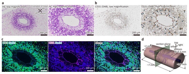
- Figure 2 Stained image of a cross-section of the tubular liver tissue perpendicular to the main blood vessel. ( a ) hematoxylin and eosin (HE) staining image. ( b ) immunohistochemistry (IHC) image (CD31). Asterisks indicate the main blood vessels. Blue arrowheads indicate capillaries. Red arrowheads indicate branches from main vessels to capillaries. ( c ) Immunofluorescence image (CK18 and CD31). ( d ) Schematic image of the tubular liver tissue.
- Submitted by
- Invitrogen Antibodies (provider)
- Main image

- Experimental details
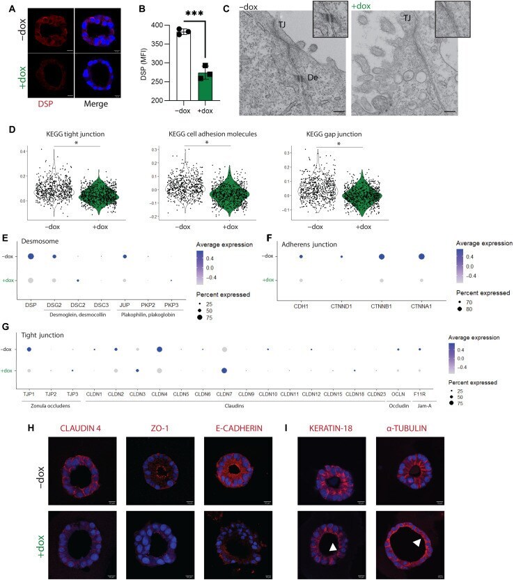
- Figure 2 Primary bEEC culture and identification. (A) Morphological observation of bEECs by ordinary optical microscopy (magnification, x200). (B) Primary bEECs were positive for the epithelial-specific marker CK-18 (red). (C) Nuclei were stained with DAPI (blue). (D) Merge of CK-18 and DAPI (magnification, x400). (E) The CK-18 + cells were counted using ImageJ. bEEC, bovine endometrial epithelial cell; CK-18, cytokeratin-18; IF, immunofluorescence.

- Submitted by
- Invitrogen Antibodies (provider)
- Main image
- Experimental details
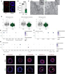
- Fig. 5. DSP -kd disrupts cell-cell junctions and cytoskeletal organization. CRISPRi-iAT2s were transduced with DSP gRNA and treated without (-dox) or with (+dox) doxycycline. ( A ) Immunofluorescence staining of desmoplakin (DSP). DSP, red; nuclei, blue; scale bar, 10 mum. ( B ) Flow cytometry analysis of DSP expression, represented by mean fluorescence intensity (MFI). n = 3 experimental replicates of independent wells of a differentiation; error bars represent SD. Statistical significance was determined by unpaired, two-tailed Student's t test; *** P < 0.001. ( C ) Transmission electron microscopy images showing tight junctions (TJ) and desmosomes (De); scale bar, 200 nm. ( D ) Violin plots showing module scores for KEGG (Kyoto Encyclopedia of Genes and Genomes) tight junction, cell adhesion molecules, and gap junction. ( E ) Dot plots showing expression of genes associated with desmosome, ( F ) adherens junction, and ( G ) TJ. Genes that were not expressed by iAT2s were excluded from dot plots. ( H ) Immunofluorescence staining of claudin-4, ZO-1, and E-cadherin. ( I ) Immunofluorescence staining of keratin-18 and alpha-tubulin. Arrows indicate disorganized filaments or tubules. Protein of interest, red; nuclei, blue; scale bar, 10 mum.
- Submitted by
- Invitrogen Antibodies (provider)
- Main image
- Experimental details
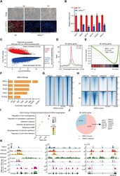
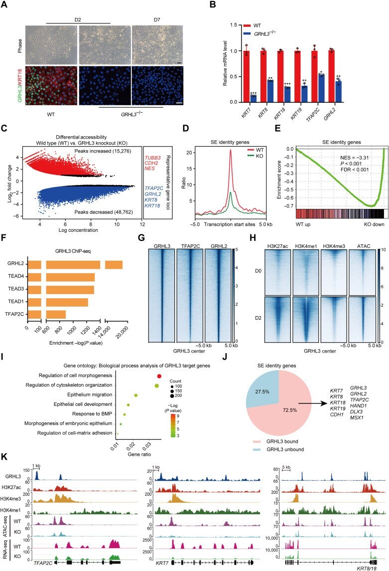
- Fig. 3. GRHL3 is required to activate SE identity genes through opening their chromatin accessibilities. ( A ) Wild-type (WT) and GRHL3 -knockout hESCs during SE differentiation. Top: Phase contrast images. Bottom: Immunofluorescence staining of GRHL3 (green) and KRT18 (red). Scale bars, 100 mum. ( B ) Quantitative reverse transcription polymerase chain reaction (qRT-PCR) analysis of representative genes in wild-type and GRHL3 -knockout hESCs after 2 days of differentiation. qRT-PCR values were normalized to the values in wild-type group. Values are presented as means +- SD ( n = 3 biological replicates; * P < 0.05; ** P < 0.01; *** P < 0.001; t test). ( C ) Scatterplot of differential accessibility in wild-type versus GRHL3 -knockout (KO) hESCs after 2 days of differentiation. Sites identified as significantly differentially bound [log 2 fold change > 1 or < -1 and false discovery rate (FDR) < 0.05] are shown in color (red, peaks increased; blue, peaks decreased). ( D ) Metaplots of average ATAC-seq density around the SE identity genes in wild-type and GRHL3 -knockout hESCs after 2 days of differentiation. ( E ) Gene set enrichment analysis of the SE identity gene set in the gene expression matrix of wild-type and GRHL3 -knockout hESCs after 2 days of differentiation. NES, normalized enrichment score. ( F ) Enrichment of transcription factor motifs identified by HOMER at GRHL3 peaks. ( G ) Heatmaps of the binding signals of TFAP2C and GRHL2 at the center of GRHL3 peaks. ( H
- Submitted by
- Invitrogen Antibodies (provider)
- Main image
- Experimental details
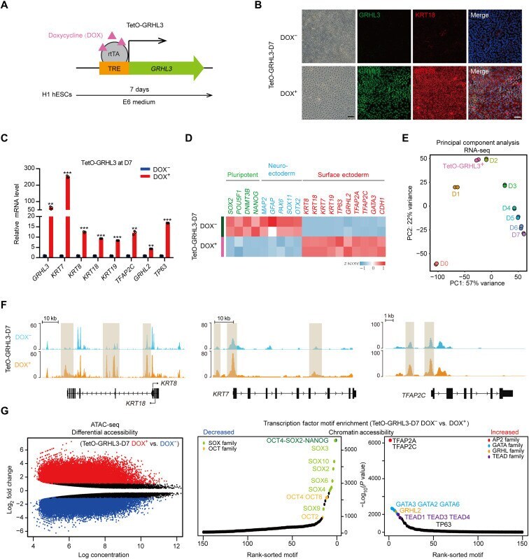
- Fig. 4. GRHL3 induces SE commitment. ( A ) Schematic diagram of establishment of a TetO-GRHL3 doxycycline-inducible expression system in hESC. Cells were induced with doxycycline for 7 days. TRE, tetracycline response element; rtTA, reverse tetracycline transactivator. ( B ) Immunofluorescence staining of GRHL3 (green) and KRT18 (red) in TetO-GRHL3 + and TetO-GRHL3 - cells. Left: Phase contrast images. Scale bars, 100 mum. ( C ) qRT-PCR analysis of representative genes in TetO-GRHL3 + and TetO-GRHL3 - cells. qRT-PCR values were normalized to the values in TetO-GRHL3 - cells. Values are presented as means +- SD ( n = 3 biological replicates; ** P < 0.01; *** P < 0.001; t test). ( D ) Heatmap of germ layer-specific gene expression levels in TetO-GRHL3 + and TetO-GRHL3 - cells. ( E ) PCA of RNA-seq data of TetO-GRHL3 + cells and hESCs at each time points during SE differentiation. ( F ) Genome browser tracks comparing ATAC-seq signal at KRT8/18, KRT7, and TFAP2C loci in TetO-GRHL3 + and TetO-GRHL3 - cells. ( G ) Left: Scatterplot of differential accessibility in TetO-GRHL3 + versus TetO-GRHL3 - cells. Sites identified as significantly differentially bound are shown in color (red, peaks increased; blue, peaks decreased). Right: Transcription factor motif enrichment in the regions of differential accessibility in TetO-GRHL3 + versus TetO-GRHL3 - cells. TEAD, TEA domain transcription factor.
- Submitted by
- Invitrogen Antibodies (provider)
- Main image
- Experimental details
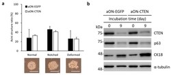
- Figure 3 Inducible EGFP-expressing and CTEN-expressing RWPE-1 cells perform similar phenotypes in the absence of doxycycline in 3D culture. Inducible EGFP-expressing (aON-EGFP) or CTEN-expressing (aON-CTEN) RWPE-1 cells were incubated in 3D culture system for 9 days without doxycycline induction. ( a ) The appearance of acinar structures was observed and classified to three types (normal, notched and deformed). The numbers of the three-type acini in each group were counted and presented as a percentage of the whole (upper panel). Data were expressed as mean +- SD of three independent experiments. More than 50 acini were evaluated within each sample (Student's t test; no significant difference between aON-EGFP and aON-CTEN in each phenotype). The representative images of acinar structure were also shown in the lower panel. Scale bar: 100 mum. ( b ) aON-EGFP and aON-CTEN RWPE-1 cells grown in 3D culture were collected at the indicated time and the protein levels were examined by Western analyses using the indicated antibodies. alpha-tubulin was used as a loading control.

- Submitted by
- Invitrogen Antibodies (provider)
- Main image
- Experimental details
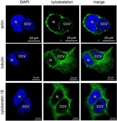
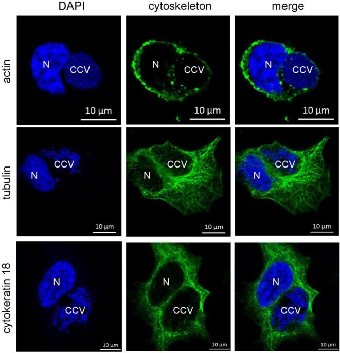
- Figure 10 Cytoskeletal filaments decorate the CCV. HeLa cells were infected with C. burnetii at an MOI of 50. At 72 h post-infection, cells were fixed and stained with Phallotoxin-647 for actin, or with tubulin- and cytokeratin 18-specific antibodies by indirect immunofluorescence (green). Nuclei and bacterial DNA were stained with DAPI (blue). Cells were visualized using LSM. N: Nucleus. CCV: C. burnetii -containing vacuole.
- Submitted by
- Invitrogen Antibodies (provider)
- Main image
- Experimental details
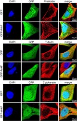
- Figure 11 Cytoskeletal filaments are not modified by AnkF expression. HeLa cells were transiently transfected with pGFP alone or with pEGFP-AnkF (green). 24 h post-transfection, cells were fixed and stained with Phalloidin-Alexa647 for actin, or with tubulin- and cytokeratin 18-specific antibodies by indirect immunofluorescence (red). Nuclei and bacterial DNA were stained with DAPI (blue). Cells were visualized using LSM. Representative images from two independent experiments are shown.
