Explore
 Validate
 Learn
Learn Western blot
Antibody data
- Antibody Data
- Antigen structure
- References [1]
- Comments [0]
- Validations
- Western blot [2]
- Immunocytochemistry [1]
- Immunohistochemistry [1]
Submit
Validation data
Reference
Comment
Report error
- Product number
- MAB7619 - Provider product page

- Provider
- R&D Systems
- Product name
- Human Cytokeratin 18 Antibody
- Antibody type
- Monoclonal
- Description
- Protein A or G purified from hybridoma culture supernatant. Detects human Cytokertin 18 in direct ELISAs and Western blots.
- Reactivity
- Human
- Host
- Mouse
- Conjugate
- Unconjugated
- Antigen sequence
P05783- Isotype
- IgG
- Antibody clone number
- 810811
- Vial size
- 100 ug
- Concentration
- LYOPH
- Storage
- Use a manual defrost freezer and avoid repeated freeze-thaw cycles. 12 months from date of receipt, -20 to -70 °C as supplied. 1 month, 2 to 8 °C under sterile conditions after reconstitution. 6 months, -20 to -70 °C under sterile conditions after reconstitution.
Submitted references TGFβ1 in fibroblasts-derived exosomes promotes epithelial-mesenchymal transition of ovarian cancer cells.
Li W, Zhang X, Wang J, Li M, Cao C, Tan J, Ma D, Gao Q
Oncotarget 2017 Nov 10;8(56):96035-96047
Oncotarget 2017 Nov 10;8(56):96035-96047
No comments: Submit comment
Supportive validation
- Submitted by
- R&D Systems (provider)
- Main image
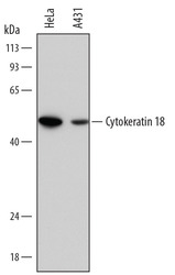
- Experimental details
- Detection of Human Cytokeratin 18 by Western Blot. Western blot shows lysates of HeLa human cervical epithelial carcinoma cell line and A431 human epithelial carcinoma cell line. PVDF membrane was probed with 0.2 µg/mL of Mouse Anti-Human Cytokeratin 18 Monoclonal Antibody (Catalog # MAB7619) followed by HRP-conjugated Anti-Mouse IgG Secondary Antibody (Catalog # HAF018). A specific band was detected for Cytokeratin 18 at approximately 48 kDa (as indicated). This experiment was conducted under reducing conditions and using Immunoblot Buffer Group 1.
- Submitted by
- R&D Systems (provider)
- Main image
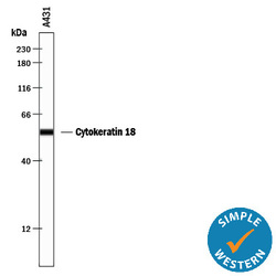
- Experimental details
- Detection of Human Cytokeratin 18 by Simple WesternTM. Simple Western lane view shows lysates of A431 human epithelial carcinoma cell line, loaded at 0.5 mg/mL. A specific band was detected for Cytokeratin 18 at approximately 55 kDa (as indicated) using 2 µg/mL of Mouse Anti-Human Cytokeratin 18 Monoclonal Antibody (Catalog # MAB7619). This experiment was conducted under reducing conditions and using the 12-230 kDa separation system.

Supportive validation
- Submitted by
- R&D Systems (provider)
- Main image
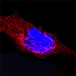
- Experimental details
- Cytokeratin 18 in HeLa Human Cell Line. Cytokeratin 18 was detected in immersion fixed HeLa human cervical epithelial carcinoma cell line using Mouse Anti-Human Cytokeratin 18 Monoclonal Antibody (Catalog # MAB7619) at 25 µg/mL for 3 hours at room temperature. Cells were stained using the NorthernLights™ 557-conjugated Anti-Mouse IgG Secondary Antibody (red; Catalog # NL007) and counterstained with DAPI (blue). Specific staining was localized to intermediate filaments in cytoplasm. View our protocol for Fluorescent ICC Staining of Cells on Coverslips.

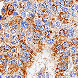
Supportive validation
- Submitted by
- R&D Systems (provider)
- Main image

- Experimental details
- Cytokeratin 18 in Human Squamous Cell Carcinoma. Cytokeratin 18 was detected in immersion fixed paraffin-embedded sections of human squamous cell carcinoma using Mouse Anti-Human Cytokeratin 18 Monoclonal Antibody (Catalog # MAB7619) at 15 µg/mL overnight at 4 °C. Tissue was stained using the Anti-Mouse HRP-DAB Cell & Tissue Staining Kit (brown; Catalog # CTS002) and counterstained with hematoxylin (blue). Specific staining was localized to cancer cells. View our protocol for Chromogenic IHC Staining of Paraffin-embedded Tissue Sections.