Explore
 Validate
 Learn
LearnPA1-41139
antibody from Invitrogen Antibodies
Targeting: TLR5
FLJ10052, MGC126430, MGC126431, SLEB1, TIL3
 Western blot
 Immunohistochemistry
Antibody data
- Antibody Data
- Antigen structure
- References [2]
- Comments [0]
- Validations
- Immunohistochemistry [1]
- Flow cytometry [3]
- Other assay [2]
Submit
Validation data
Reference
Comment
Report error
- Product number
- PA1-41139 - Provider product page

- Provider
- Invitrogen Antibodies
- Product name
- TLR5 Polyclonal Antibody
- Antibody type
- Polyclonal
- Antigen
- Synthetic peptide
- Reactivity
- Human, Mouse, Rat, Porcine
- Host
- Rabbit
- Isotype
- IgG
- Vial size
- 100 μg
- Concentration
- 1.0 mg/mL
- Storage
- -20°C, Avoid Freeze/Thaw Cycles
Submitted references TLR5 participates in the TLR4 receptor complex and promotes MyD88-dependent signaling in environmental lung injury.
Flagellin/TLR5 signalling activates renal collecting duct cells and facilitates invasion and cellular translocation of uropathogenic Escherichia coli.
Hussain S, Johnson CG, Sciurba J, Meng X, Stober VP, Liu C, Cyphert-Daly JM, Bulek K, Qian W, Solis A, Sakamachi Y, Trempus CS, Aloor JJ, Gowdy KM, Foster WM, Hollingsworth JW, Tighe RM, Li X, Fessler MB, Garantziotis S
eLife 2020 Jan 28;9
eLife 2020 Jan 28;9
Flagellin/TLR5 signalling activates renal collecting duct cells and facilitates invasion and cellular translocation of uropathogenic Escherichia coli.
Bens M, Vimont S, Ben Mkaddem S, Chassin C, Goujon JM, Balloy V, Chignard M, Werts C, Vandewalle A
Cellular microbiology 2014 Oct;16(10):1503-17
Cellular microbiology 2014 Oct;16(10):1503-17
No comments: Submit comment
Supportive validation
- Submitted by
- Invitrogen Antibodies (provider)
- Main image
- Experimental details
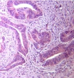
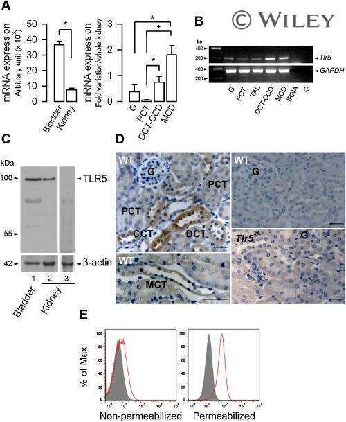
- Immunohistochemical analysis of TLR5 in formalin-fixed paraffin-embedded human stomach tumor tissue. Samples were incubated in TLR5 polyclonal antibody (Product # PA1-41139) using a dilution of 10 µg/mL.

Supportive validation
- Submitted by
- Invitrogen Antibodies (provider)
- Main image

- Experimental details
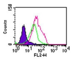
- Intracellular flow analysis of TLR5 in Balb/c mouse splenocytes using a TLR5 polyclonal antibody (Product # PA1-41139) at 2 µg/10^6 cells. Shaded histogram represents cells without antibody; green represents rabbit IgG isotype control; red represents TLR5 polyclonal antibody (Product # PA1-41139). Goat anti-rabbit PE was used as secondary antibody.

- Submitted by
- Invitrogen Antibodies (provider)
- Main image

- Experimental details
- Flow cytometry of TLR5 in Balb/c mouse splenocytes. Samples were incubated in TLR5 polyclonal antibody (Product # PA1-41139) using a dilution of 2 µg/10^6 cells followed by Goat anti-rabbit PE secondary antibody. Shaded histogram: cells without antibody. Green: rabbit IgG isotype control. Red: TLR5 antibody.

- Submitted by
- Invitrogen Antibodies (provider)
- Main image
- Experimental details
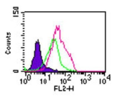
- Flow cytometry of TLR5 in Balb/c mouse splenocytes. Samples were incubated in TLR5 polyclonal antibody (Product # PA1-41139) using a dilution of 2 µg/10^6 cells followed by Goat anti-rabbit PE secondary antibody. Shaded histogram: cells without antibody. Green: rabbit IgG isotype control. Red: TLR5 antibody.
Supportive validation
- Submitted by
- Invitrogen Antibodies (provider)
- Main image
- Experimental details
- NULL
- Submitted by
- Invitrogen Antibodies (provider)
- Main image

- Experimental details
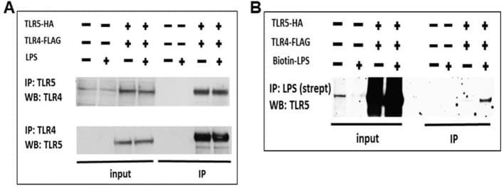
- Figure 8. TLR5 participates in TLR4 signaling complex. ( A ) Co-immunoprecipitation of hemagglutinin-tagged TLR5 (TLR5-HA) and FLAG-tagged TLR4 (TLR4-FLAG) in HEK293 cells after 100 ng/mL ultrapure LPS exposure. ( B ) Immunoprecipitation of TLR5 with biotinylated ultrapure LPS (Biotin-LPS) in TLR5-HA and TLR4-FLAG transfected HEK293 cells after 100 ng/mL Biotin-LPS exposure for 15 min. Representative of 2 separate experiments. Figure 8--figure supplement 1. Cell surface expression of TLR4 and its co-localization with TLR5.