Explore
 Validate
 Learn
Learn Western blot
Antibody data
- Antibody Data
- Antigen structure
- References [2]
- Comments [0]
- Validations
- Western blot [7]
- Immunohistochemistry [2]
Submit
Validation data
Reference
Comment
Report error
- Product number
- GTX124127 - Provider product page

- Provider
- GeneTex
- Proper citation
- GeneTex Cat#GTX124127, RRID:AB_11170703
- Product name
- DLK/MAP3K12 antibody
- Antibody type
- Polyclonal
- Reactivity
- Human, Mouse, Rat
- Host
- Rabbit
Submitted references The Ste20 Family Kinases MAP4K4, MINK1, and TNIK Converge to Regulate Stress-Induced JNK Signaling in Neurons.
Akt suppresses DLK for maintaining self-renewal of mouse embryonic stem cells.
Larhammar M, Huntwork-Rodriguez S, Rudhard Y, Sengupta-Ghosh A, Lewcock JW
The Journal of neuroscience : the official journal of the Society for Neuroscience 2017 Nov 15;37(46):11074-11084
The Journal of neuroscience : the official journal of the Society for Neuroscience 2017 Nov 15;37(46):11074-11084
Akt suppresses DLK for maintaining self-renewal of mouse embryonic stem cells.
Wu CC, Wu HJ, Wang CH, Lin CH, Hsu SC, Chen YR, Hsiao M, Schuyler SC, Lu FL, Ma N, Lu J
Cell cycle (Georgetown, Tex.) 2015;14(8):1207-17
Cell cycle (Georgetown, Tex.) 2015;14(8):1207-17
No comments: Submit comment
Supportive validation
- Submitted by
- GeneTex (provider)
- Main image
- Experimental details
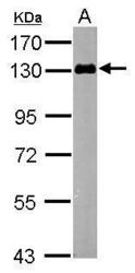
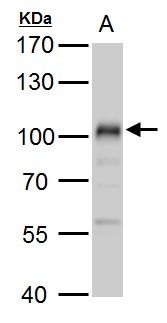
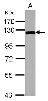
- Sample (50 ?g of whole cell lysate) A: mouse brain 7.5% SDS PAGE GTX124127 diluted at 1:1000 The HRP-conjugated anti-rabbit IgG antibody (GTX213110-01) was used to detect the primary antibody.

- Submitted by
- GeneTex (provider)
- Main image

- Experimental details
- Sample (20 ?g of whole cell lysate) A: mouse ESC 7.5% SDS PAGE GTX124127 diluted at 1:500 The HRP-conjugated anti-rabbit IgG antibody (GTX213110-01) was used to detect the primary antibody.
- Submitted by
- GeneTex (provider)
- Main image
- Experimental details
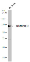
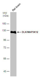
- DLK/MAP3K12 antibody detects DLK/MAP3K12 protein by western blot analysis.A. 50 ?g rat brain lysate/extract7.5 % SDS-PAGEDLK/MAP3K12 antibody (GTX124127) dilution: 1:1000
- Validation comment
- WB
- Submitted by
- GeneTex (provider)
- Main image
- Experimental details
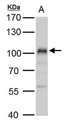
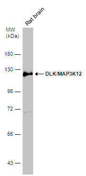
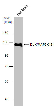
- Rat tissue extract (50 ?g) was separated by 7.5% SDS-PAGE, and the membrane was blotted with DLK/MAP3K12 antibody (GTX124127) diluted at 1:1000. The HRP-conjugated anti-rabbit IgG antibody (GTX213110-01) was used to detect the primary antibody.
- Submitted by
- GeneTex (provider)
- Main image

- Experimental details
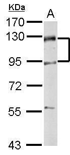
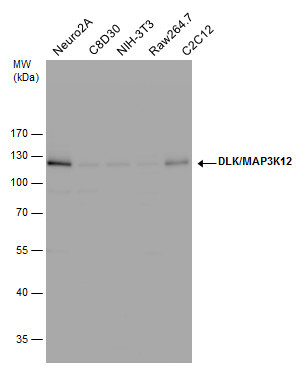
- DLK/MAP3K12 antibody detects DLK/MAP3K12 protein by western blot analysis. Various whole cell extracts (30 ?g) were separated by 10% SDS-PAGE, and the membrane was blotted with DLK/MAP3K12 antibody (GTX124127) diluted by 1:1000. The HRP-conjugated anti-rabbit IgG antibody (GTX213110-01) was used to detect the primary antibody.
- Submitted by
- GeneTex (provider)
- Main image
- Experimental details
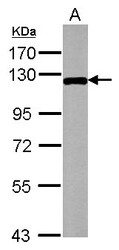
- Rat tissue extract (50 ?g) was separated by 7.5% SDS-PAGE, and the membrane was blotted with DLK/MAP3K12 antibody (GTX124127) diluted at 1:1000.
- Submitted by
- GeneTex (provider)
- Main image
- Experimental details
- DLK/MAP3K12 antibody detects MAP3K12 protein by Western blot analysis.A. 20 £gg mouse embryonic stem cell whole cell lysate/extract 7.5 % SDS-PAGEDLK/MAP3K12 antibody (GTX124127) dilution: 1:1000
Supportive validation
- Submitted by
- GeneTex (provider)
- Main image
- Experimental details
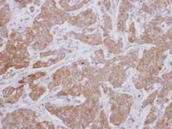
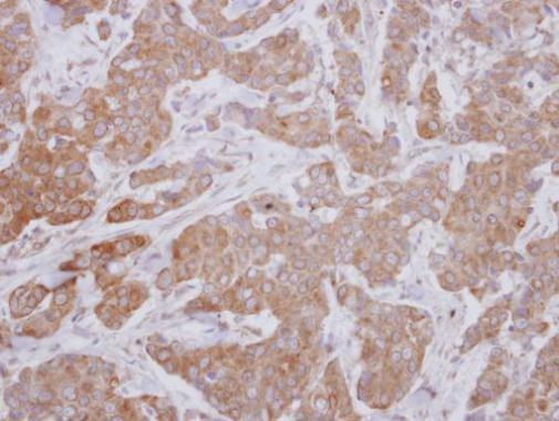
- Immunohistochemical analysis of paraffin-embedded human breast cancer, using MAP3K12(GTX124127) antibody at 1:250 dilution.
- Submitted by
- GeneTex (provider)
- Main image
- Experimental details
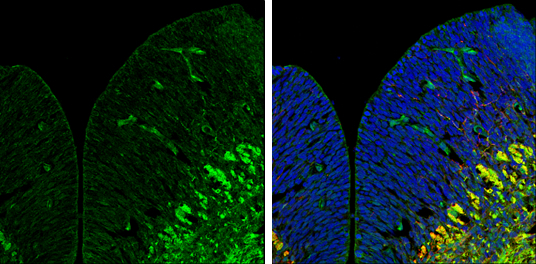
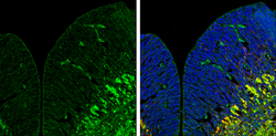
- DLK/MAP3K12 antibody detects DLK/MAP3K12 protein expression by immunohistochemical analysis.Sample: Frozen sectioned E13.5 Rat brain. Green: DLK/MAP3K12 protein stained by DLK/MAP3K12 antibody (GTX124127) diluted at 1:250.Red: beta Tubulin 3/ TUJ1, a mature neuron marker, stained by beta Tubulin 3/ TUJ1 antibody [GT11710] (GTX631836) diluted at 1:500.Blue: Fluoroshield with DAPI (GTX30920).