Explore
 Validate
 Learn
Learn Western blot
 Immunohistochemistry
Antibody data
- Antibody Data
- Antigen structure
- References [3]
- Comments [0]
- Validations
- Immunohistochemistry [1]
Submit
Validation data
Reference
Comment
Report error
- Product number
- AF2254 - Provider product page

- Provider
- R&D Systems
- Product name
- Mouse DNER Antibody
- Antibody type
- Polyclonal
- Description
- Antigen Affinity-purified. Detects mouse DNER in direct ELISAs and Western blots. In direct ELISAs and Western blots, approximately 40% cross-reactivity with recombinant human DNER is observed.
- Reactivity
- Mouse
- Host
- Goat
- Conjugate
- Unconjugated
- Antigen sequence
Q8JZM4- Isotype
- IgG
- Vial size
- 100 ug
- Concentration
- LYOPH
- Storage
- Use a manual defrost freezer and avoid repeated freeze-thaw cycles. 12 months from date of receipt, -20 to -70 °C as supplied. 1 month, 2 to 8 °C under sterile conditions after reconstitution. 6 months, -20 to -70 °C under sterile conditions after reconstitution.
Submitted references The Notch ligand DNER regulates macrophage IFNγ release in chronic obstructive pulmonary disease.
Delta/Notch-Like EGF-Related Receptor (DNER) Is Not a Notch Ligand.
A search for factors specifying tonotopy implicates DNER in hair-cell development in the chick's cochlea.
Ballester-López C, Conlon TM, Ertüz Z, Greiffo FR, Irmler M, Verleden SE, Beckers J, Fernandez IE, Eickelberg O, Yildirim AÖ
EBioMedicine 2019 May;43:562-575
EBioMedicine 2019 May;43:562-575
Delta/Notch-Like EGF-Related Receptor (DNER) Is Not a Notch Ligand.
Greene M, Lai Y, Pajcini K, Bailis W, Pear WS, Lancaster E
PloS one 2016;11(9):e0161157
PloS one 2016;11(9):e0161157
A search for factors specifying tonotopy implicates DNER in hair-cell development in the chick's cochlea.
Kowalik L, Hudspeth AJ
Developmental biology 2011 Jun 15;354(2):221-31
Developmental biology 2011 Jun 15;354(2):221-31
No comments: Submit comment
Supportive validation
- Submitted by
- R&D Systems (provider)
- Main image
- Experimental details
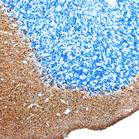
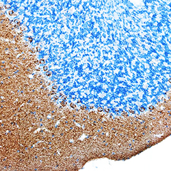
- DNER in Mouse Brain. DNER was detected in perfusion fixed frozen sections of mouse brain (cerebellum) using Goat Anti-Mouse DNER Antigen Affinity-purified Polyclonal Antibody (Catalog # AF2254) at 5 µg/mL overnight at 4 °C. Tissue was stained using the Anti-Goat HRP-DAB Cell & Tissue Staining Kit (brown; Catalog # CTS008) and counterstained with hematoxylin (blue). Specific staining was localized to Purkinje neurons and molecular layer. View our protocol for Chromogenic IHC Staining of Frozen Tissue Sections.