Explore
 Validate
 Learn
Learn Western blot
 Immunocytochemistry
Antibody data
- Antibody Data
- Antigen structure
- References [2]
- Comments [0]
- Validations
- Immunocytochemistry [3]
- Flow cytometry [2]
- Other assay [2]
Submit
Validation data
Reference
Comment
Report error
- Product number
- 459120 - Provider product page

- Provider
- Invitrogen Antibodies
- Product name
- COX5A Monoclonal Antibody (6E9B12D5)
- Antibody type
- Monoclonal
- Antigen
- Other
- Description
- Positive control: Isolated mitochondria from Human heart, Bovine heart, Rat heart, Mouse heart, and HepG2, Human embryonic lung-derived fibroblasts (strain MRC5), HeLa cells.
- Reactivity
- Human, Mouse, Rat, Bovine
- Host
- Mouse
- Isotype
- IgG
- Antibody clone number
- 6E9B12D5
- Vial size
- 100 μg
- Concentration
- 1 mg/mL
- Storage
- 4°C, do not freeze
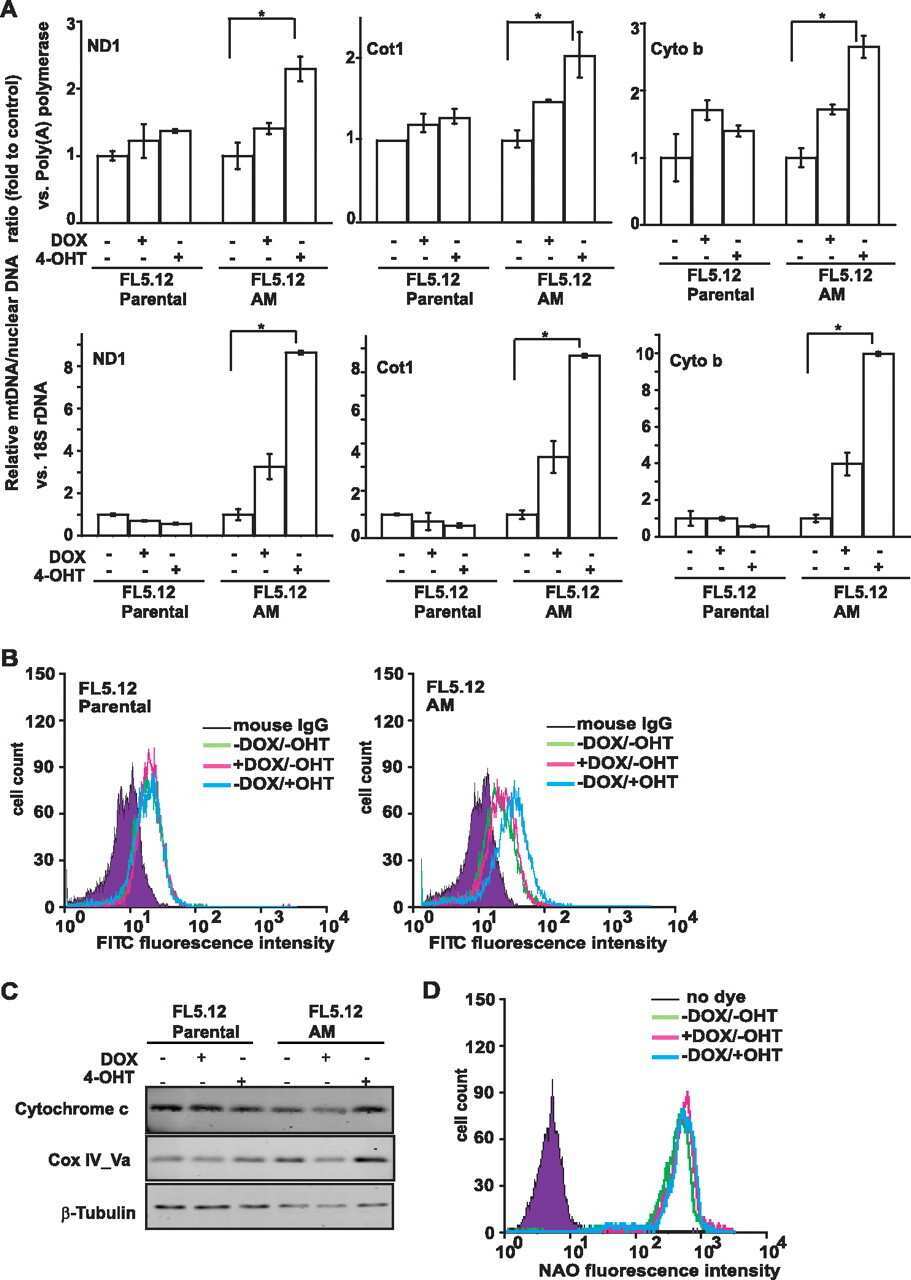
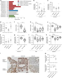
Submitted references Ulcerative colitis mucosal transcriptomes reveal mitochondriopathy and personalized mechanisms underlying disease severity and treatment response.
Akt and c-Myc differentially activate cellular metabolic programs and prime cells to bioenergetic inhibition.
Haberman Y, Karns R, Dexheimer PJ, Schirmer M, Somekh J, Jurickova I, Braun T, Novak E, Bauman L, Collins MH, Mo A, Rosen MJ, Bonkowski E, Gotman N, Marquis A, Nistel M, Rufo PA, Baker SS, Sauer CG, Markowitz J, Pfefferkorn MD, Rosh JR, Boyle BM, Mack DR, Baldassano RN, Shah S, Leleiko NS, Heyman MB, Grifiths AM, Patel AS, Noe JD, Aronow BJ, Kugathasan S, Walters TD, Gibson G, Thomas SD, Mollen K, Shen-Orr S, Huttenhower C, Xavier RJ, Hyams JS, Denson LA
Nature communications 2019 Jan 3;10(1):38
Nature communications 2019 Jan 3;10(1):38
Akt and c-Myc differentially activate cellular metabolic programs and prime cells to bioenergetic inhibition.
Fan Y, Dickman KG, Zong WX
The Journal of biological chemistry 2010 Mar 5;285(10):7324-33
The Journal of biological chemistry 2010 Mar 5;285(10):7324-33
No comments: Submit comment
Supportive validation
- Submitted by
- Invitrogen Antibodies (provider)
- Main image

- Experimental details
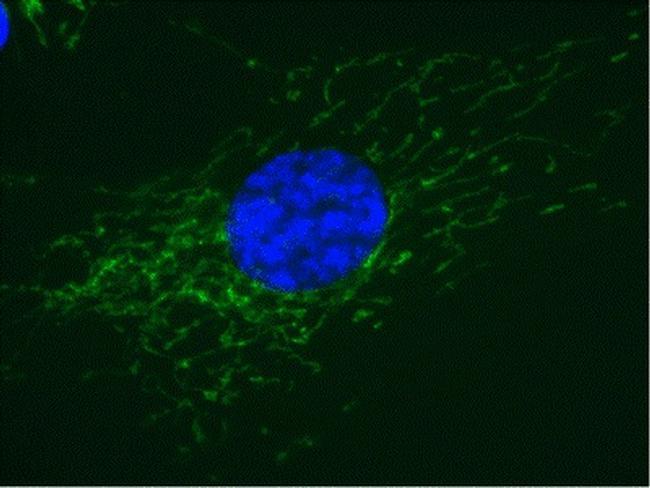
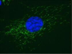
- Immunofluorescent analysis of Complex IV in MRC5 cells using a Complex IV Monoclonal antibody (Product # 459120) at a concentration of 10 µg/mL. Cultured Human embryonic lung-derived fibroblasts (strain MRC5) were fixed, treated for heat-induced antigen retrieval, permeabilized, and then labeled with Complex IV Monoclonal antibody (Product # 459120) at a concentration of 10 µg/mL. An AlexaFluor® 488-conjugated-goat-anti-mouse IgG2a isotype specific secondary antibody at a concentration of 1 µg/mL was used for detection.
- Submitted by
- Invitrogen Antibodies (provider)
- Main image
- Experimental details
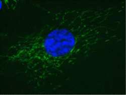
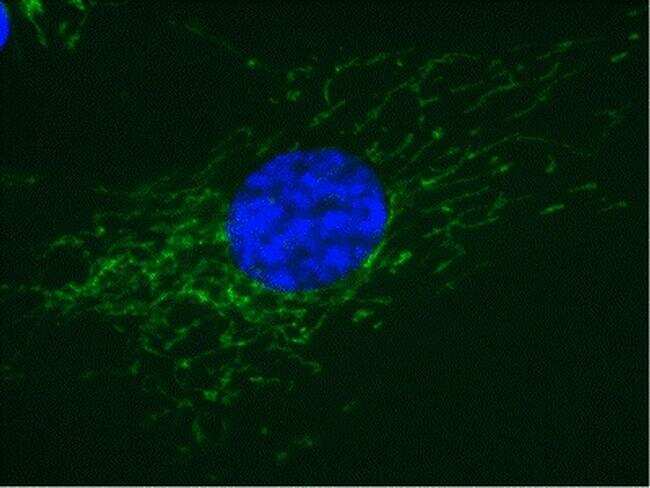
- Immunofluorescent analysis of Complex IV in MRC5 cells using a Complex IV Monoclonal antibody (Product # 459120) at a concentration of 10 µg/mL. Cultured Human embryonic lung-derived fibroblasts (strain MRC5) were fixed, treated for heat-induced antigen retrieval, permeabilized, and then labeled with Complex IV Monoclonal antibody (Product # 459120) at a concentration of 10 µg/mL. An AlexaFluor® 488-conjugated-goat-anti-mouse IgG2a isotype specific secondary antibody at a concentration of 1 µg/mL was used for detection.

Supportive validation
- Submitted by
- Invitrogen Antibodies (provider)
- Main image

- Experimental details
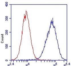
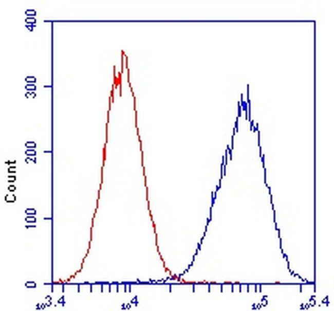
- Flow cytometric analysis of Complex IV in HeLa cells using a Complex IV monoclonal antibody (Product # 459120) at 1µg/mL is depicted by the blue line. The red line indicates an isotype control antibody.

- Submitted by
- Invitrogen Antibodies (provider)
- Main image
- Experimental details
- Flow cytometric analysis of Complex IV in HeLa cells using a Complex IV monoclonal antibody (Product # 459120) at 1µg/mL is depicted by the blue line. The red line indicates an isotype control antibody.
Supportive validation
- Submitted by
- Invitrogen Antibodies (provider)
- Main image
- Experimental details
- NULL
- Submitted by
- Invitrogen Antibodies (provider)
- Main image
- Experimental details
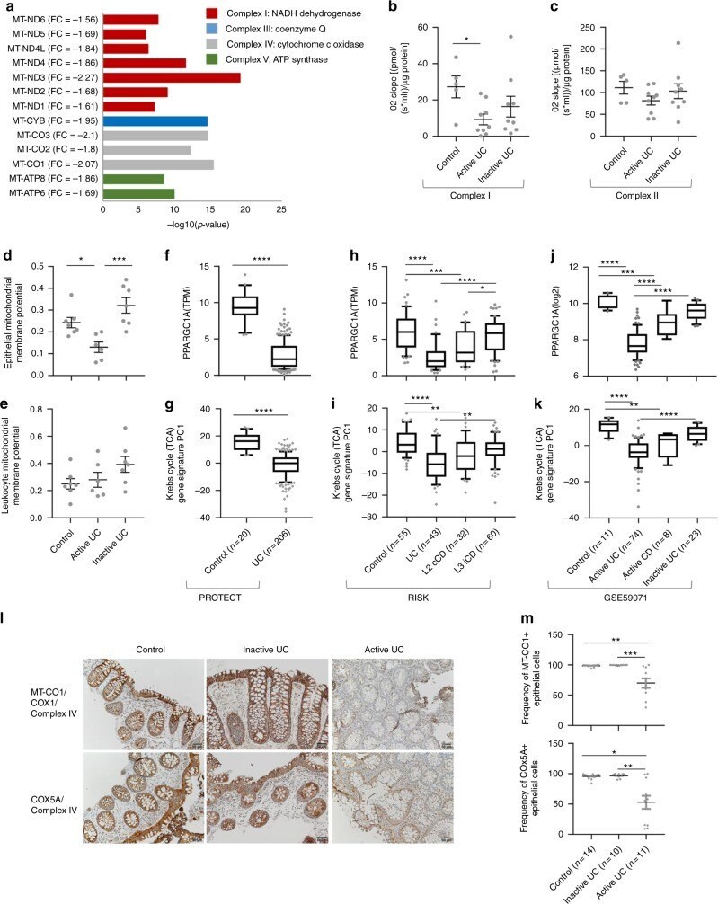
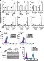
- Fig. 2 Colonic mitochondriopathy with a robust gene signature for reduced rectal mitochondrial energy functions in UC. a Thirteen mitochondrial-encoded genes are downregulated in UC vs. control with their fold change, FDR corrected P value, and associated mitochondrial complex as indicated. High-Resolution Respirometry was performed on fresh colon biopsies (5 control, 9 with active UC, and 9 with inactive UC) using the Oroboros O2k modular system to evaluate the activity of Complex I ( b ) and Complex II ( c ) of the electron transport chain. JC1 staining and FACS analysis were used to define the mitochondrial membrane potential of d EpCAM + epithelial cells and e CD45 + leukocytes isolated from colon biopsies (7 controls, 6 active UC, and 7 with inactive UC, 85-99% viability). Colon PPARGC1A (PGC-1alpha) expression for f PROTECT cohort, h RISK cohort in (transcripts per million (TPM) values), and for j adult UC cohort (GSE59071 12 ) in normalized values was plotted after stratifying the samples as indicated. g , i , k Krebs cycle TCA gene signature PCA PC1 for the above cohorts is plotted, samples are stratified as indicated. l Representative rectal MT-CO1 and COX5A immunohistochemistry (complex IV) for Ctl ( n = 14), inactive ( n = 10), and active UC ( n = 11) with moderate Mayo endoscopic subscore and moderate PUCAI. Scale bar represents 50 mum. m Frequency of MT-CO1-positive and COX5A-positive epithelial cells out of the total epithelial cells for