 Explore
 Validate
 Learn
Learn Western blot
 ELISA
ELISA Immunocytochemistry
Antibody data
- Antibody Data
- Antigen structure
- References [11]
- Comments [0]
- Validations
- Immunocytochemistry [2]
- Other assay [5]
Submit
Validation data
Reference
Comment
Report error
- Product number
- AMC3012 - Provider product page

- Provider
- Invitrogen Antibodies
- Product name
- TNF alpha Polyclonal Antibody
- Antibody type
- Polyclonal
- Antigen
- Recombinant full-length protein
- Description
- Reconstitute using 0.5 mL sterile distilled water.
- Reactivity
- Human, Mouse
- Host
- Rabbit
- Vial size
- 500 μg
- Concentration
- 1 mg/mL
- Storage
- 4°C
Submitted references Isolation of DiNP-Degrading Microbes from the Mouse Colon and the Influence DiNP Exposure Has on the Microbiota, Intestinal Integrity, and Immune Status of the Colon.
Glycine protects partial liver grafts from Kupffer cell-dependent ischemia-reperfusion injury without negative effect on regeneration.
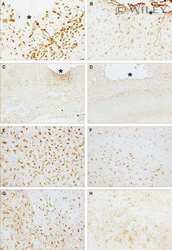
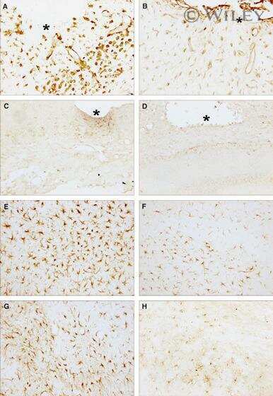
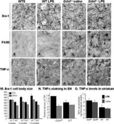
Prenatal LPS increases inflammation in the substantia nigra of Gdnf heterozygous mice.
Metallic silver fragments cause massive tissue loss in the mouse brain.
Focal cerebral ischemia in the TNFalpha-transgenic rat.
Elevated levels of IL-18 in plasma and skeletal muscle in chronic obstructive pulmonary disease.
Fiber type specific expression of TNF-alpha, IL-6 and IL-18 in human skeletal muscles.
Metallothionein reduces central nervous system inflammation, neurodegeneration, and cell death following kainic acid-induced epileptic seizures.
A critical role for alveolar macrophages in elicitation of pulmonary immune fibrosis.
An IFN-gamma-independent proinflammatory role of IL-18 in murine streptococcal cell wall arthritis.
Enhancement of antigen-presenting cell surface molecules involved in cognate interactions by immunostimulatory DNA sequences.
Chiu KK, Bashir ST, Abdel-Hamid AM, Clark LV, Laws MJ, Cann I, Nowak RA, Flaws JA
Toxics 2022 Feb 6;10(2)
Toxics 2022 Feb 6;10(2)
Glycine protects partial liver grafts from Kupffer cell-dependent ischemia-reperfusion injury without negative effect on regeneration.
Al-Saeedi M, Liang R, Schultze DP, Nickkholgh A, Herr I, Zorn M, Schemmer P
Amino acids 2019 Jun;51(6):903-911
Amino acids 2019 Jun;51(6):903-911
Prenatal LPS increases inflammation in the substantia nigra of Gdnf heterozygous mice.
Granholm AC, Zaman V, Godbee J, Smith M, Ramadan R, Umphlet C, Randall P, Bhat NR, Rohrer B, Middaugh LD, Boger HA
Brain pathology (Zurich, Switzerland) 2011 May;21(3):330-48
Brain pathology (Zurich, Switzerland) 2011 May;21(3):330-48
Metallic silver fragments cause massive tissue loss in the mouse brain.
Locht LJ, Pedersen MØ, Markholt S, Bibby BM, Larsen A, Penkowa M, Stoltenberg M, Rungby J
Basic & clinical pharmacology & toxicology 2011 Jul;109(1):1-10
Basic & clinical pharmacology & toxicology 2011 Jul;109(1):1-10
Focal cerebral ischemia in the TNFalpha-transgenic rat.
Pettigrew LC, Kindy MS, Scheff S, Springer JE, Kryscio RJ, Li Y, Grass DS
Journal of neuroinflammation 2008 Oct 23;5:47
Journal of neuroinflammation 2008 Oct 23;5:47
Elevated levels of IL-18 in plasma and skeletal muscle in chronic obstructive pulmonary disease.
Petersen AM, Penkowa M, Iversen M, Frydelund-Larsen L, Andersen JL, Mortensen J, Lange P, Pedersen BK
Lung 2007 May-Jun;185(3):161-71
Lung 2007 May-Jun;185(3):161-71
Fiber type specific expression of TNF-alpha, IL-6 and IL-18 in human skeletal muscles.
Plomgaard P, Penkowa M, Pedersen BK
Exercise immunology review 2005;11:53-63
Exercise immunology review 2005;11:53-63
Metallothionein reduces central nervous system inflammation, neurodegeneration, and cell death following kainic acid-induced epileptic seizures.
Penkowa M, Florit S, Giralt M, Quintana A, Molinero A, Carrasco J, Hidalgo J
Journal of neuroscience research 2005 Feb 15;79(4):522-34
Journal of neuroscience research 2005 Feb 15;79(4):522-34
A critical role for alveolar macrophages in elicitation of pulmonary immune fibrosis.
Zhang-Hoover J, Sutton A, van Rooijen N, Stein-Streilein J
Immunology 2000 Dec;101(4):501-11
Immunology 2000 Dec;101(4):501-11
An IFN-gamma-independent proinflammatory role of IL-18 in murine streptococcal cell wall arthritis.
Joosten LA, van De Loo FA, Lubberts E, Helsen MM, Netea MG, van Der Meer JW, Dinarello CA, van Den Berg WB
Journal of immunology (Baltimore, Md. : 1950) 2000 Dec 1;165(11):6553-8
Journal of immunology (Baltimore, Md. : 1950) 2000 Dec 1;165(11):6553-8
Enhancement of antigen-presenting cell surface molecules involved in cognate interactions by immunostimulatory DNA sequences.
Martin-Orozco E, Kobayashi H, Van Uden J, Nguyen MD, Kornbluth RS, Raz E
International immunology 1999 Jul;11(7):1111-8
International immunology 1999 Jul;11(7):1111-8
No comments: Submit comment
Supportive validation
- Submitted by
- Invitrogen Antibodies (provider)
- Main image

- Experimental details
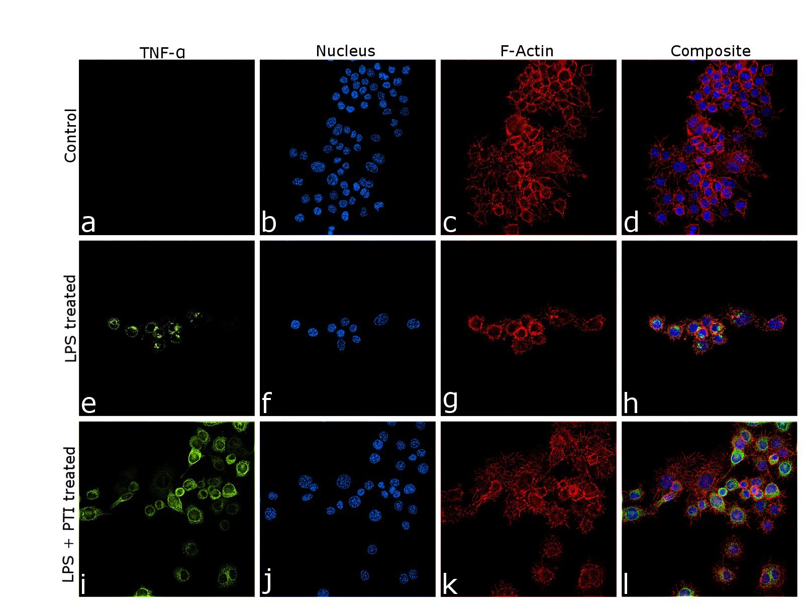
- Immunofluorescence analysis of TNF-Alpha (Tumor necrosis factor alpha) was performed using 70% confluent log phase RAW 264.7 treated with lipopolysaccharide (LPS) (10 ng/mL for 7 hr) and protein transport inhibitor cocktail [500X] (PTI) (1X for 4 hr). The cells were fixed with 4% paraformaldehyde for 10 minutes, permeabilized with 0.1% Triton™ X-100 for 10 minutes and blocked with 2% BSA for 45 minutes at room temperature. The cells were labeled with TNF alpha Polyclonal Antibody (Product # AMC3012) at 1:100 in 0.1% BSA, incubated at 4 degree celsius overnight and then labeled with Donkey anti-Rabbit IgG (H+L) Highly Cross-Adsorbed Secondary Antibody, Alexa Fluor Plus 488 (Product # A32790), (1:2500), for 45 minutes at room temperature (Panel a,e,i: Green). Nuclei (Panel b,f,j: Blue) were stained with ProLong™ Diamond Antifade Mountant with DAPI (Product # P36962). F-actin (Panel c,g,k: Red) was stained with Rhodamine Phalloidin (Product # R415, 1:300). Panel d represents the merged image of untreated cells showing no staining for TNF-alpha and panel h and i shows upregulation of TNF-alpha expression in golgi-like structures upon LPS stimulation only and intracellular signal accumulation upon subsequent treatment with the secretory blocker PTI, respectively. The images were captured at 60X magnification.
- Submitted by
- Invitrogen Antibodies (provider)
- Main image

- Experimental details
- Immunofluorescence analysis of TNF-Alpha (Tumor necrosis factor alpha) was performed using 70% confluent log phase RAW 264.7 treated with lipopolysaccharide (LPS) (10 ng/mL for 7 hr) and protein transport inhibitor cocktail [500X] (PTI) (1X for 4 hr). The cells were fixed with 4% paraformaldehyde for 10 minutes, permeabilized with 0.1% Triton™ X-100 for 10 minutes and blocked with 2% BSA for 45 minutes at room temperature. The cells were labeled with TNF alpha Polyclonal Antibody (Product # AMC3012) at 1:100 in 0.1% BSA, incubated at 4 degree celsius overnight and then labeled with Donkey anti-Rabbit IgG (H+L) Highly Cross-Adsorbed Secondary Antibody, Alexa Fluor Plus 488 (Product # A32790), (1:2500), for 45 minutes at room temperature (Panel a,e,i: Green). Nuclei (Panel b,f,j: Blue) were stained with ProLong™ Diamond Antifade Mountant with DAPI (Product # P36962). F-actin (Panel c,g,k: Red) was stained with Rhodamine Phalloidin (Product # R415, 1:300). Panel d represents the merged image of untreated cells showing no staining for TNF-alpha and panel h and i shows upregulation of TNF-alpha expression in golgi-like structures upon LPS stimulation only and intracellular signal accumulation upon subsequent treatment with the secretory blocker PTI, respectively. The images were captured at 60X magnification.

Supportive validation
- Submitted by
- Invitrogen Antibodies (provider)
- Main image

- Experimental details
- NULL
- Submitted by
- Invitrogen Antibodies (provider)
- Main image
- Experimental details
- NULL
- Submitted by
- Invitrogen Antibodies (provider)
- Main image
- Experimental details
- NULL
- Submitted by
- Invitrogen Antibodies (provider)
- Main image
- Experimental details
- NULL

- Submitted by
- Invitrogen Antibodies (provider)
- Main image
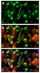
- Experimental details
- Figure 3 Identification of neural cell type containing TNFalpha protein . This photographic collage shows the result of co-localization studies identifying the neural cell type containing TNFalpha protein in the brain of the transgenic animal. All images were taken of TNFalpha-transgenic rat brain that had not been subjected to ischemic injury. Coronal brain sections were incubated with primary antibodies identifying mouse or rat TNFalpha protein and neurons. All images were made by fluorescence microscopy at 40x magnification. In Panel a, cortical neurons in TNFalpha-Tg rat brain are immuno-labeled with the anti-neuronal antibody NeuN tagged with a green fluorophore. In Panel b, co-localization images are merged to show that several NeuN-labeled neurons (green) express TNFalpha protein immuno-labeled with anti-mouse/rat TNFalpha antibody tagged with a red fluorophore. In Panel c, co-localization images are merged to show NeuN-labeled neurons (green), TNFalpha protein (red), and cellular nuclei labeled with Hoechst stain (blue). A representative neuron with cytoplasmic inclusions labeled to show expression of TNFalpha protein is identified in each Panel (arrow). In Panel c, nuclei of several non-neuronal cells that do not express TNFalpha protein are shown. Scale bar = 50 mum
