Explore
 Validate
 Learn
Learn Western blot
Antibody data
- Antibody Data
- Antigen structure
- References [2]
- Comments [0]
- Validations
- Western blot [1]
- Immunohistochemistry [1]
Submit
Validation data
Reference
Comment
Report error
- Product number
- sc-14021 - Provider product page

- Provider
- Santa Cruz Biotechnology
- Proper citation
- Santa Cruz Biotechnology Cat#sc-14021, RRID:AB_2174944
- Product name
- Anti-PTPN1
- Antibody type
- Polyclonal
- Antigen
- Recombinant full-length protein
- Reactivity
- Human
- Host
- Rabbit
Submitted references Inhibition of PTP1B restores IRS1-mediated hepatic insulin signaling in IRS2-deficient mice.
Silencing of SH-PTP2 defines a crucial role in the inactivation of epidermal growth factor receptor by 5-aminosalicylic acid in colon cancer cells
González-Rodríguez A, Mas Gutierrez JA, Sanz-González S, Ros M, Burks DJ, Valverde AM
Diabetes 2010 Mar;59(3):588-99
Diabetes 2010 Mar;59(3):588-99
Silencing of SH-PTP2 defines a crucial role in the inactivation of epidermal growth factor receptor by 5-aminosalicylic acid in colon cancer cells
G Monteleone, L Franchi, D Fina, R Caruso, P Vavassori, I Monteleone, E Calabrese, G C Naccari, S Bellinvia, R Testi, F Pallone
Cell Death and Differentiation 2005 Aug;13(2):202-211
Cell Death and Differentiation 2005 Aug;13(2):202-211
No comments: Submit comment
Supportive validation
- Submitted by
- per
- Main image

- Experimental details
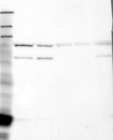
- Western blot analysis of antibody specificity using a routine panel composed of IgG/HSA-depleted human plasma and protein lysates from selected human tissues and cell lines.
- Validation comment
- Band of predicted size in kDa (+/-20%) with additional bands present.
- Primary Ab dilution
- 1:500
- Secondary Ab dilution
- 1:3000
- Lane 1
- Marker [kDa]: 219, 111, 83, 48, 32, 26, 17
- Lane 2
- RT-4
- Lane 3
- U-251MG sp
- Lane 4
- Human Plasma
- Lane 5
- Liver
- Lane 6
- Tonsil
- Theoretical target weight
- [kDa] 50
Supportive validation
- Submitted by
- per
- Main image

- Experimental details
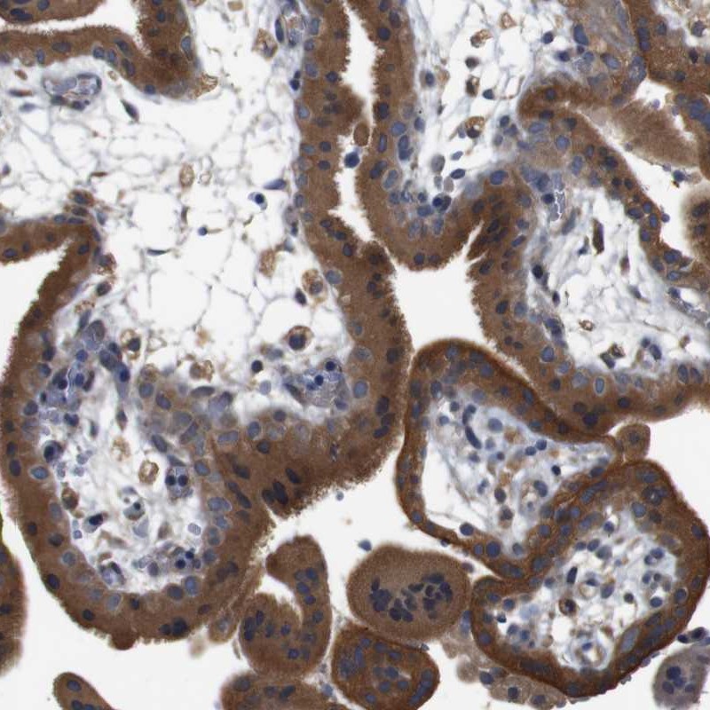
- Immunohistochemical staining of human placenta shows strong cytoplasmic positivity in trophoblastic cells.
- Validation comment
- Staining pattern consistent with experimental and/or bioinformatic data.