 Explore
 Validate
 Learn
Learn Western blot
Antibody data
- Antibody Data
- Antigen structure
- References [6]
- Comments [0]
- Validations
- Western blot [3]
- Immunohistochemistry [1]
Submit
Validation data
Reference
Comment
Report error
- Product number
- AF1366 - Provider product page

- Provider
- Novus Biologicals
- Product name
- Rabbit Polyclonal PTP1B/PTPN1 Antibody
- Antibody type
- Polyclonal
- Description
- Antigen Affinity-purified. Detects endogenous human PTP1B in Western blots.
- Reactivity
- Human
- Host
- Rabbit
- Conjugate
- Unconjugated
- Isotype
- IgG
- Vial size
- 100 ug
- Concentration
- LYOPH
- Storage
- Use a manual defrost freezer and avoid repeated freeze-thaw cycles. 12 months from date of receipt, -20 to -70 degreesC as supplied. 1 month, 2 to 8 degreesC under sterile conditions after reconstitution. 6 months, -20 to -70 degreesC under sterile conditions after reconstitution.
Submitted references The pseudophosphatase phogrin enables glucose-stimulated insulin signaling in pancreatic β cells.
Inhibition of PTP1B disrupts cell-cell adhesion and induces anoikis in breast epithelial cells.
Selective activation of SHP2 activity by cisplatin revealed by a novel chemical probe-based assay.
Calpain 2 and PTP1B function in a novel pathway with Src to regulate invadopodia dynamics and breast cancer cell invasion.
Cysteine S-nitrosylation protects protein-tyrosine phosphatase 1B against oxidation-induced permanent inactivation.
Protein cysteine sulfinic acid reductase (sulfiredoxin) as a regulator of cell proliferation and drug response.
Torii S, Kubota C, Saito N, Kawano A, Hou N, Kobayashi M, Torii R, Hosaka M, Kitamura T, Takeuchi T, Gomi H
The Journal of biological chemistry 2018 Apr 20;293(16):5920-5933
The Journal of biological chemistry 2018 Apr 20;293(16):5920-5933
Inhibition of PTP1B disrupts cell-cell adhesion and induces anoikis in breast epithelial cells.
Hilmarsdottir B, Briem E, Halldorsson S, Kricker J, Ingthorsson S, Gustafsdottir S, Mælandsmo GM, Magnusson MK, Gudjonsson T
Cell death & disease 2017 May 11;8(5):e2769
Cell death & disease 2017 May 11;8(5):e2769
Selective activation of SHP2 activity by cisplatin revealed by a novel chemical probe-based assay.
Kuo CC, Chu CY, Lin JJ, Lo LC
Biochemical and biophysical research communications 2010 Jan 1;391(1):230-4
Biochemical and biophysical research communications 2010 Jan 1;391(1):230-4
Calpain 2 and PTP1B function in a novel pathway with Src to regulate invadopodia dynamics and breast cancer cell invasion.
Cortesio CL, Chan KT, Perrin BJ, Burton NO, Zhang S, Zhang ZY, Huttenlocher A
The Journal of cell biology 2008 Mar 10;180(5):957-71
The Journal of cell biology 2008 Mar 10;180(5):957-71
Cysteine S-nitrosylation protects protein-tyrosine phosphatase 1B against oxidation-induced permanent inactivation.
Chen YY, Chu HM, Pan KT, Teng CH, Wang DL, Wang AH, Khoo KH, Meng TC
The Journal of biological chemistry 2008 Dec 12;283(50):35265-72
The Journal of biological chemistry 2008 Dec 12;283(50):35265-72
Protein cysteine sulfinic acid reductase (sulfiredoxin) as a regulator of cell proliferation and drug response.
Lei K, Townsend DM, Tew KD
Oncogene 2008 Aug 21;27(36):4877-87
Oncogene 2008 Aug 21;27(36):4877-87
No comments: Submit comment
Supportive validation
- Submitted by
- Novus Biologicals (provider)
- Main image

- Experimental details
- Detection of Human PTP1B by Western Blot. Western blot shows lysates of HeLa human cervical epithelial carcinoma cell line and TF-1 human erythroleukemic cell line. PVDF membrane was probed with 1 µg/mL of Rabbit Anti-Human PTP1B Antigen Affinity-purified Polyclonal Antibody (Catalog # AF1366) followed by HRP-conjugated Anti-Rabbit IgG Secondary Antibody (Catalog # HAF008). A specific band was detected for PTP1B at approximately 50 kDa (as indicated). This experiment was conducted under reducing conditions and using Immunoblot Buffer Group 1.

- Submitted by
- Novus Biologicals (provider)
- Main image

- Experimental details
- Detection of Human PTP1B by Simple WesternTM. Simple Western lane view shows lysates of HeLa human cervical epithelial carcinoma cell line, loaded at 0.2 mg/mL. A specific band was detected for PTP1B at approximately 57 kDa (as indicated) using 10 µg/mL of Rabbit Anti-Human PTP1B Antigen Affinity-purified Polyclonal Antibody (Catalog # AF1366). This experiment was conducted under reducing conditions and using the 12-230 kDa separation system.

- Submitted by
- Novus Biologicals (provider)
- Main image
- Experimental details
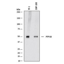
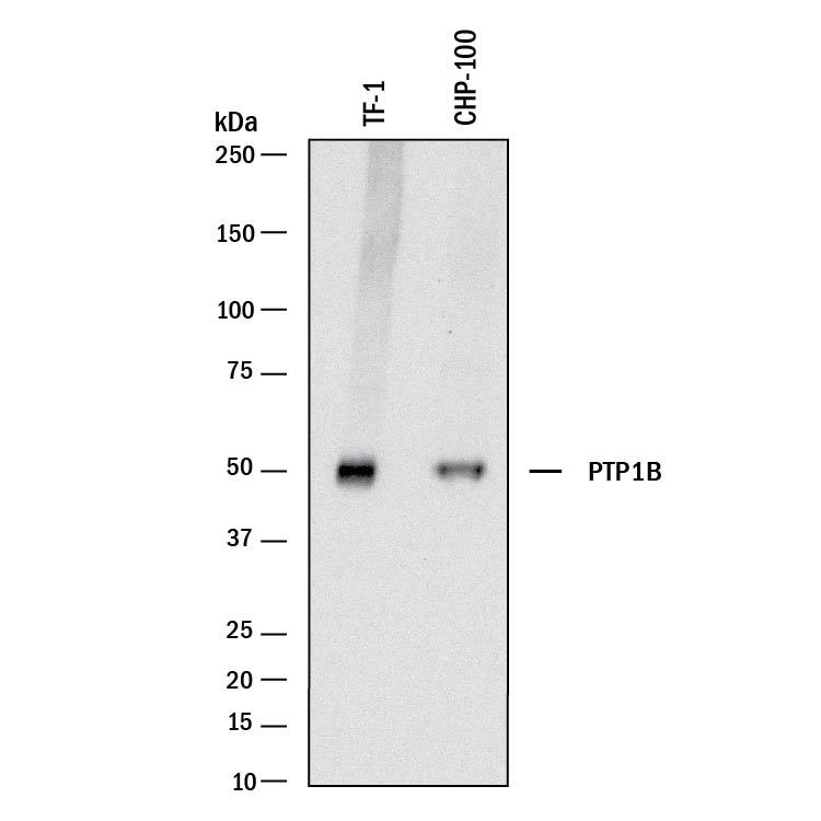
- Detection of Human PTP1B by Western Blot. Detection of Human PTP1B by Western Blot.
Supportive validation
- Submitted by
- Novus Biologicals (provider)
- Main image
- Experimental details
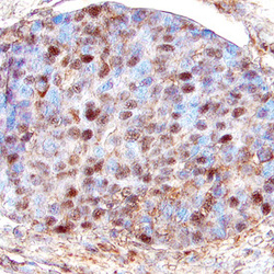
- PTP1B in Human Breast Cancer Tissue. PTP1B was detected in immersion fixed paraffin-embedded sections of human breast cancer tissue using Rabbit Anti-Human PTP1B Antigen Affinity-purified Polyclonal Antibody (Catalog # AF1366) at 15 µg/mL overnight at 4 °C. Tissue was stained using the Anti-Rabbit HRP-DAB Cell & Tissue Staining Kit (brown; Catalog # CTS005) and counterstained with hematoxylin (blue). Specific labeling was localized to the nuclei in glandular epithelial cells. View our protocol for Chromogenic IHC Staining of Paraffin-embedded Tissue Sections.
