 Explore
 Validate
 Learn
Learn Western blot
Antibody data
- Antibody Data
- Antigen structure
- References [0]
- Comments [0]
- Validations
- Western blot [4]
Submit
Validation data
Reference
Comment
Report error
- Product number
- AF13661 - Provider product page

- Provider
- R&D Systems
- Product name
- Human/Mouse/Rat PTP1B Antibody
- Antibody type
- Polyclonal
- Description
- Antigen Affinity-purified. Detects human, mouse and rat PTP1B in Western blots.
- Reactivity
- Human, Mouse, Rat
- Host
- Goat
- Conjugate
- Unconjugated
- Antigen sequence
P18031- Isotype
- IgG
- Vial size
- 100 ug
- Concentration
- LYOPH
- Storage
- Use a manual defrost freezer and avoid repeated freeze-thaw cycles. 12 months from date of receipt, -20 to -70 °C as supplied. 1 month, 2 to 8 °C under sterile conditions after reconstitution. 6 months, -20 to -70 °C under sterile conditions after reconstitution.
No comments: Submit comment
Supportive validation
- Submitted by
- R&D Systems (provider)
- Main image

- Experimental details
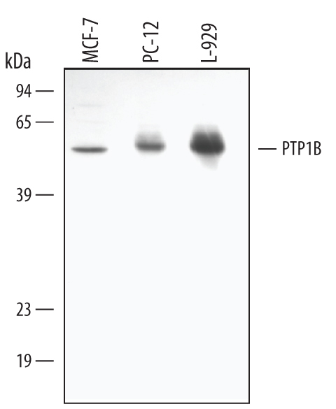
- Detection of Human/Mouse/Rat PTP1B by Western Blot. Western blot shows lysates of MCF-7 human breast cancer cell line, PC-12 rat adrenal pheochromocytoma cell line, and L-929 mouse fibroblast cell line. PVDF membrane was probed with 1 µg/mL of Goat Anti-Human/Mouse/Rat PTP1B Antigen Affinity-purified Polyclonal Antibody (Catalog # AF13661) followed by HRP-conjugated Anti-Goat IgG Secondary Antibody (Catalog # HAF109). A specific band was detected for PTP1B at approximately 50 kDa (as indicated). This experiment was conducted using Immunoblot Buffer Group 1.
- Submitted by
- R&D Systems (provider)
- Main image

- Experimental details
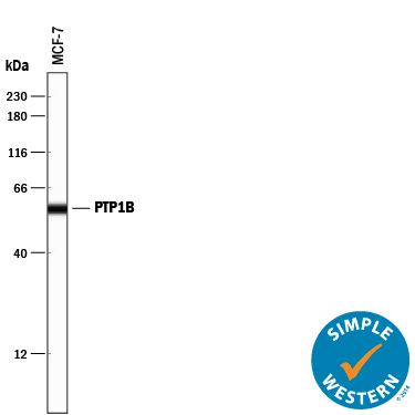
- Detection of Human PTP1B by Simple WesternTM. Simple Western lane view shows lysates of MCF-7 human breast cancer cell line, loaded at 0.2 mg/mL. A specific band was detected for PTP1B at approximately 57 kDa (as indicated) using 10 µg/mL of Goat Anti-Human/Mouse/Rat PTP1B Antigen Affinity-purified Polyclonal Antibody (Catalog # AF13661) followed by 1:50 dilution of HRP-conjugated Anti-Goat IgG Secondary Antibody (Catalog # HAF109). This experiment was conducted under reducing conditions and using the 12-230 kDa separation system.
- Submitted by
- R&D Systems (provider)
- Main image

- Experimental details
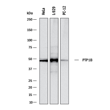
- Detection of Human, Mouse, and Rat PTP1B by Western Blot. Western blot shows lysates of HeLa human cervical epithelial carcinoma cell line, L-929 mouse fibroblast cell line, and PC-12 rat adrenal pheochromocytoma cell line. PVDF membrane was probed with 1 µg/mL of Goat Anti-Human/Mouse/Rat PTP1B Antigen Affinity-purified Polyclonal Antibody (Catalog # AF13661) followed by HRP-conjugated Anti-Goat IgG Secondary Antibody (Catalog # HAF017). A specific band was detected for PTP1B at approximately 50 kDa (as indicated). This experiment was conducted under reducing conditions and using Immunoblot Buffer Group 1.
- Submitted by
- R&D Systems (provider)
- Main image

- Experimental details
- Detection of Human PTP1B by Western Blot. Western blot shows lysates of TF-1 human erythroleukemic cell line and CHP-100 human neuroblastoma cell line. PVDF membrane was probed with 0.5 µg/mL of Goat Anti-Human/Mouse/Rat PTP1B Antigen Affinity-purified Polyclonal Antibody (Catalog # AF13661) followed by HRP-conjugated Anti-Goat IgG Secondary Antibody (Catalog # HAF017). A specific band was detected for PTP1B at approximately 50 kDa (as indicated). This experiment was conducted under reducing conditions and using Immunoblot Buffer Group 1.
